Copyright
©The Author(s) 2015.
World J Gastroenterol. Apr 7, 2015; 21(13): 4089-4095
Published online Apr 7, 2015. doi: 10.3748/wjg.v21.i13.4089
Published online Apr 7, 2015. doi: 10.3748/wjg.v21.i13.4089
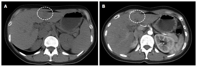
Figure 1 Computed tomography of lymphoepithelioma-like cholangiocarcinoma (dash circle).
A: Noncontrast CT showed a small hypodense nodule (38 HU) at the lateral segment of liver; B: The tumor nodule depicted enhancement in early arterial phase of contrast enhanced CT (65HU).
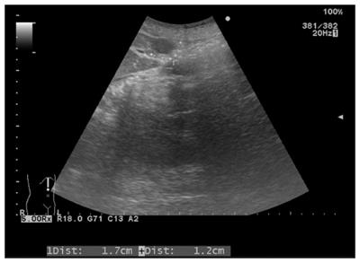
Figure 2 Sonography of lymphoepithelioma-like cholangiocarcinoma.
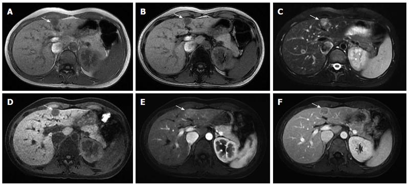
Figure 3 Liver magnetic resonance images demonstrated a small lymphoepithelioma-like cholangiocarcinoma at the medial segment of the liver (arrow).
A: In-phase T1-weighted image; B: Out of phase T1-weighted image; C: Axial T2-weighted image with fat suppression; D: Pre-contrast; E: Arterial T1-weighted image; F: Portal venous phase T1-weighted image with fat suppression.
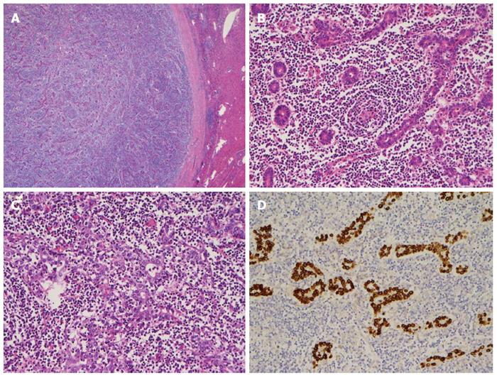
Figure 4 Histochemical findings.
A: The tumor is well circumscribed with a thick fibrous capsule (HE stain, × 20); B: The majority of the tumor is composed of well differentiated glandular cells with dense lymphocytic infiltration and scattered lymphoid follicles (HE stain, × 200); high-power view of another area in the tumor; C: The tumor reveals a solid, poorly differentiated growth pattern (HE stain, × 200); D: In situ hybridization for Epstein-Barr virus-encoded RNA is diffusely positive in the tumor cells (× 200).
- Citation: Liao TC, Liu CA, Chiu NC, Yeh YC, Chiou YY. Lymphoepithelioma-like cholangiocarcinoma: A mimic of hepatocellular carcinoma on imaging features. World J Gastroenterol 2015; 21(13): 4089-4095
- URL: https://www.wjgnet.com/1007-9327/full/v21/i13/4089.htm
- DOI: https://dx.doi.org/10.3748/wjg.v21.i13.4089


