Copyright
©The Author(s) 2015.
World J Gastroenterol. Apr 7, 2015; 21(13): 4030-4037
Published online Apr 7, 2015. doi: 10.3748/wjg.v21.i13.4030
Published online Apr 7, 2015. doi: 10.3748/wjg.v21.i13.4030
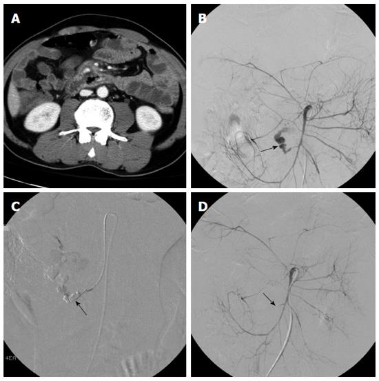
Figure 1 Images from a 36-year-old woman with a history of abdominal trauma (hemoglobin, 7.
1 g/dL; hematocrit, 24.5%). A: 64-row computed tomography angiogram in arterial phase showed a focus of extravasation in the ileum (arrow); B: Active bleeding site was found on conventional angiography; C, D: Post-embolization angiography confirmed complete occlusion of the active gastrointestinal bleeding with coils (arrow).
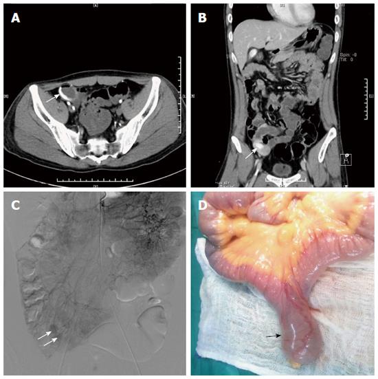
Figure 2 Images from a 64-year-old man with acute ileal bleeding (hemoglobin, 7.
4 g/dL; hematocrit, 23.7%). A, B: Axial and coronal computed tomography angiograms in arterial phase showed a focus of active extravasation (arrow) at the distal ileum; C: Superior mesenteric angiography prior to surgical resection confirmed the active extravasation (arrow); D: Emergency surgery was required 2 d after bleeding, and the resected specimen confirmed a diverticulum (arrow) at the distal ileum.
- Citation: Ren JZ, Zhang MF, Rong AM, Fang XJ, Zhang K, Huang GH, Chen PF, Wang ZY, Duan XH, Han XW, Liu YJ. Lower gastrointestinal bleeding: Role of 64-row computed tomographic angiography in diagnosis and therapeutic planning. World J Gastroenterol 2015; 21(13): 4030-4037
- URL: https://www.wjgnet.com/1007-9327/full/v21/i13/4030.htm
- DOI: https://dx.doi.org/10.3748/wjg.v21.i13.4030


