Copyright
©The Author(s) 2015.
World J Gastroenterol. Jan 7, 2015; 21(1): 60-83
Published online Jan 7, 2015. doi: 10.3748/wjg.v21.i1.60
Published online Jan 7, 2015. doi: 10.3748/wjg.v21.i1.60
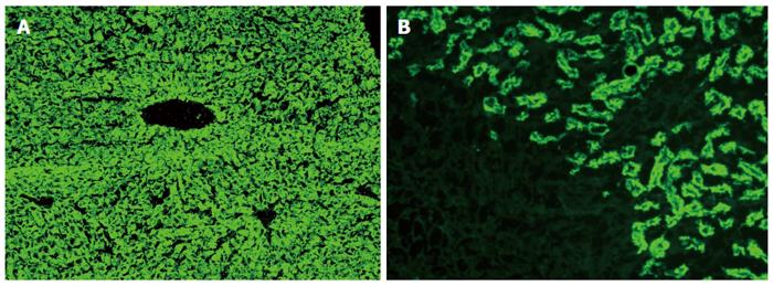
Figure 1 Typical diffuse staining of the cytoplasm of the entire liver lobule on rat liver sections in a case of autoimmune hepatitis type 2 positive for antibodies against liver-kidney microsomes type 1 (A); Antibodies against liver-kidney microsomes type 1 react to the proximal tubules of the rat kidney and the absence of reactivity against the distal tubules of the rat kidney (see also Figure 2) and parietal cells of the rat stomach distinguishes anti-LKM1 autoantibodies from antimitochondrial antibodies (B).
Original magnification × 40.
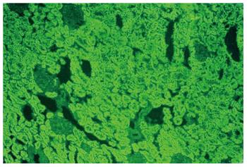
Figure 2 Contrary to antibodies against liver-kidney microsomes type 1 which react to the proximal tubules, antimitochondrial antibodies react to the proximal and also the distal tubules of the rat kidney (original magnification × 40).
In these cases there is also reactivity to the parietal cells of the rat stomach.
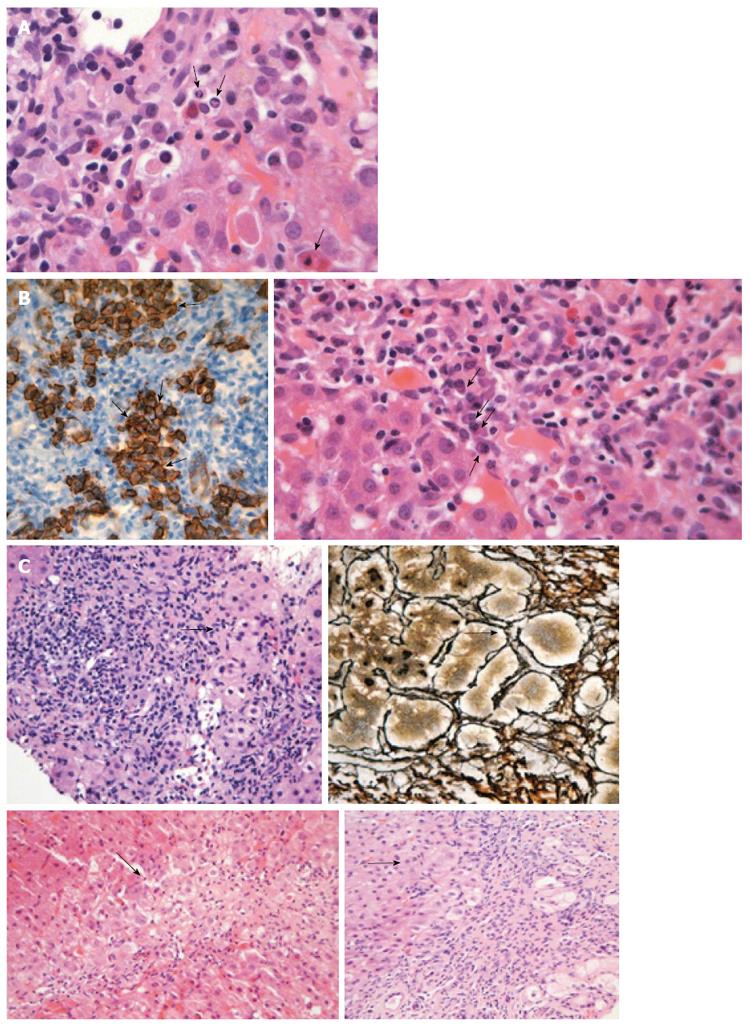
Figure 3 Liver histology in autoimmune hepatitis.
A: Interface hepatitis with genuine apoptotic bodies (arrows) in a case with autoimmune hepatitis-type 1 (hematoxylin and eosin staining). See also the typical rim of condensed chromatin at the nuclear membrane and ordinary acidophilic bodies, at various stages of development; B: Intense portal plasmacytosis in autoimmune hepatitis (arrows in the right panel; hematoxylin and eosin staining). The plasma cells are in clusters, better seen after immunostaining with CD138 (arrows in dark brown staining of the left panel). The intensity of plasmacytosis can be useful in discriminating autoimmune hepatitis from viral hepatitis whereas there is also possible prognostic information as portal plasma cell infiltrates while on immunosuppression are associated with relapse upon drug cessation; C: Various examples of rosette formation (hematoxylin and eosin staining; periportal, periseptal in a previous collapse and with reticulin staining seen in the upper right panel; see arrows).
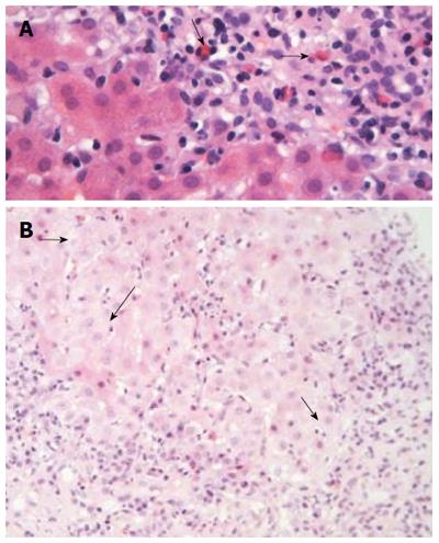
Figure 4 Liver histology in autoimmune hepatitis (continued).
A: In some cases of autoimmune hepatitis, “numerous” eosinophils or eosinophils as a minor component of the inflammatory infiltrate can be seen (arrows; hematoxylin and eosin staining). This is an interesting and useful observation, since this finding is evaluated falsely out of the histopathological context of autoimmune hepatitis, and therefore, could make the differential diagnosis from drug-induced hepatitis more problematic; B: Interface hepatitis with hydropic change of involved hepatocytes (arrows; hematoxylin and eosin staining). Note the presence of adjacent parenchymal collapse, alternatively multiacinar necrosis, which has been considered as one of the most distinguishing morphological features of autoimmune hepatitis since its presence in the appropriate clinicopathological context is supportive of the diagnosis.
- Citation: Gatselis NK, Zachou K, Koukoulis GK, Dalekos GN. Autoimmune hepatitis, one disease with many faces: Etiopathogenetic, clinico-laboratory and histological characteristics. World J Gastroenterol 2015; 21(1): 60-83
- URL: https://www.wjgnet.com/1007-9327/full/v21/i1/60.htm
- DOI: https://dx.doi.org/10.3748/wjg.v21.i1.60


