Copyright
©2014 Baishideng Publishing Group Co.
World J Gastroenterol. Mar 7, 2014; 20(9): 2176-2185
Published online Mar 7, 2014. doi: 10.3748/wjg.v20.i9.2176
Published online Mar 7, 2014. doi: 10.3748/wjg.v20.i9.2176
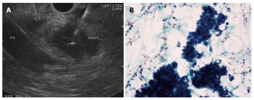
Figure 1 A 25-G needle is used to aspirate a 2 cm pancreatic head mass that does not appear to encase the portal vein (A), adenocarcinoma confirmed on wet smears obtained from the first pass in the case above (B).
Papanicolaou stain, × 40.
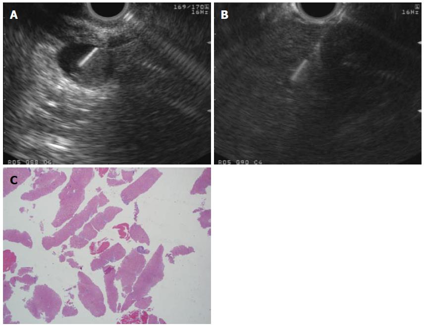
Figure 2 Flexible 19-G needle.
A: 2 cm rectal subepithelial lesion was found to originate from the muscularis propria on endoscopic ultrasound guided and is sampled using a flexible 19-G needle in this figure; B: A core liver biopsy was obtained using a flexible 19-G needle is a patient with elevated transaminases; C: Histopathological assessment of the core biopsy obtained in the case above confirmed steatohepatitis without significant fibrosis. Adequate histopathology sample was obtained and stained positively for CD-117, confirming gastrointestinal stromal tumor; H and E stain, × 2.
- Citation: Karadsheh Z, Al-Haddad M. Endoscopic ultrasound guided fine needle tissue acquisition: Where we stand in 2013? World J Gastroenterol 2014; 20(9): 2176-2185
- URL: https://www.wjgnet.com/1007-9327/full/v20/i9/2176.htm
- DOI: https://dx.doi.org/10.3748/wjg.v20.i9.2176


