Copyright
©2014 Baishideng Publishing Group Co.
World J Gastroenterol. Feb 28, 2014; 20(8): 2117-2119
Published online Feb 28, 2014. doi: 10.3748/wjg.v20.i8.2117
Published online Feb 28, 2014. doi: 10.3748/wjg.v20.i8.2117
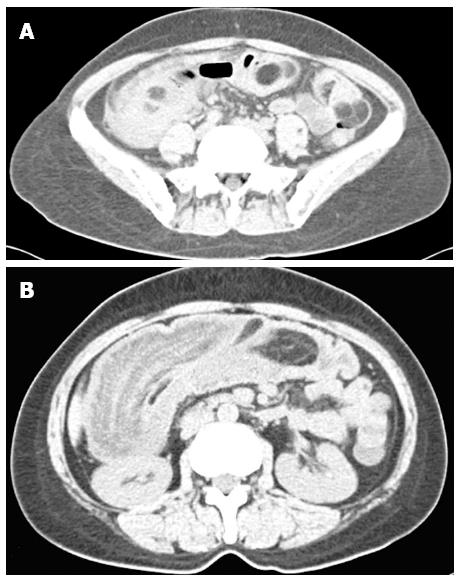
Figure 1 Abdominal computed tomography.
A: Abdominal computed tomography (CT) (transverse view) revealed diffuse and multiple intramural fat density masses in the small intestine; B: Abdominal CT showed ileo-colonic intussusception.
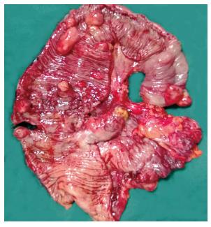
Figure 2 Multiple lipomas can be seen in the gross specimen.
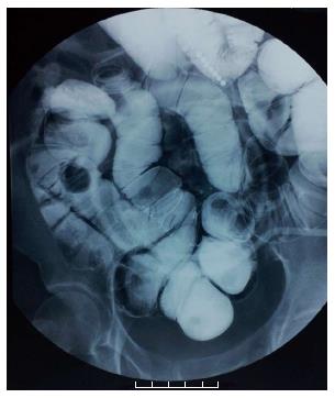
Figure 3 Small intestinal double contrast radiography revealed multiple submucosal masses in the small intestine.
- Citation: Gao PJ, Chen L, Wang FS, Zhu JY. Ileo-colonic intussusception secondary to small-bowel lipomatosis: A case report. World J Gastroenterol 2014; 20(8): 2117-2119
- URL: https://www.wjgnet.com/1007-9327/full/v20/i8/2117.htm
- DOI: https://dx.doi.org/10.3748/wjg.v20.i8.2117


