Copyright
©2014 Baishideng Publishing Group Inc.
World J Gastroenterol. Dec 21, 2014; 20(47): 18044-18047
Published online Dec 21, 2014. doi: 10.3748/wjg.v20.i47.18044
Published online Dec 21, 2014. doi: 10.3748/wjg.v20.i47.18044
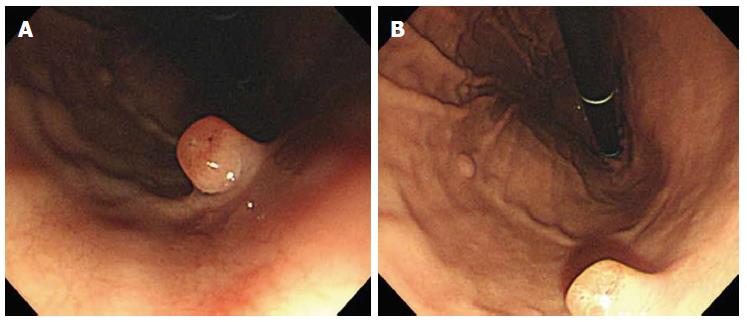
Figure 1 Endoscopic appearance of fundic gland polyps.
A: The first polyp (0.8 cm in diameter, pedunculated, reddish, and with an erosive surface) at the mid body on the anterior wall; B: Other polyps (with a smooth surface) at the mid to high body on the greater curvature.
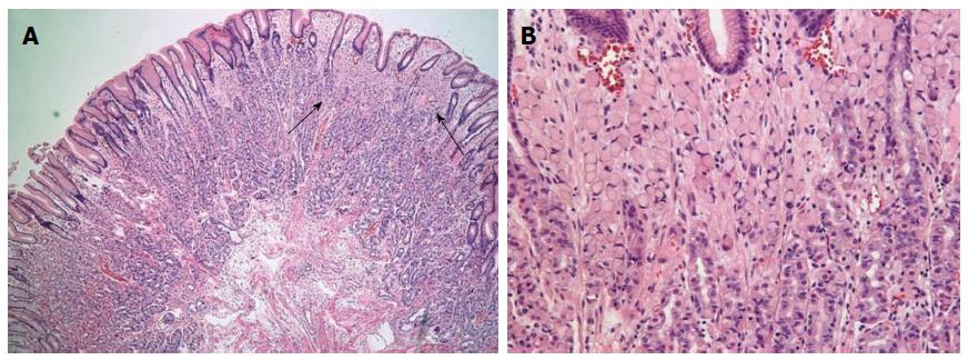
Figure 2 Histological findings.
A: A resected specimen demonstrated signet ring cell carcinoma (between the arrows) (HE staining, × 40); B: Loosely cohesive signet ring cells from fundic oxyntic glands of fundic gland polyp infiltrate the lamina propria (HE staining, × 100).
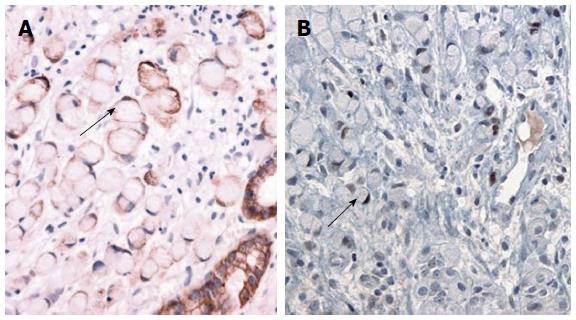
Figure 3 Immunohistochemical findings.
A: Signet ring cell carcinoma is positive for cytokeratin (arrow); B: p53 is expressed in signet ring cell carcinoma but not in the fundic oxyntic cells (arrow).
- Citation: Jeong YS, Kim SE, Kwon MJ, Seo JY, Lim H, Park JW, Kang HS, Moon SH, Kim JH, Park CK. Signet-ring cell carcinoma arising from a fundic gland polyp in the stomach. World J Gastroenterol 2014; 20(47): 18044-18047
- URL: https://www.wjgnet.com/1007-9327/full/v20/i47/18044.htm
- DOI: https://dx.doi.org/10.3748/wjg.v20.i47.18044


