Copyright
©2014 Baishideng Publishing Group Inc.
World J Gastroenterol. Dec 21, 2014; 20(47): 17993-18000
Published online Dec 21, 2014. doi: 10.3748/wjg.v20.i47.17993
Published online Dec 21, 2014. doi: 10.3748/wjg.v20.i47.17993
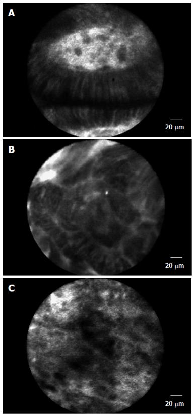
Figure 1 Probe-based confocal laser endomicroscopy images of Barrett’s esophagus.
A: Barrett‘s esophagus with intestinal metaplasia; B: Barrett‘s esophagus with dysplasia; C: Barrett‘s esophagus with neoplasia or carcinoma.
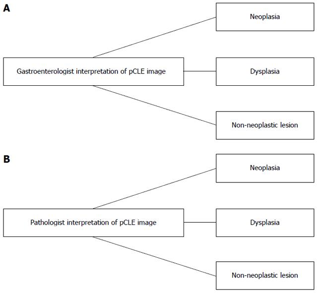
Figure 2 Flow chart for interpretation of results.
A: Anatomical site; B: Interpretation of Cellvizio image. pCLE: Probe-based confocal laser endomicroscopy.
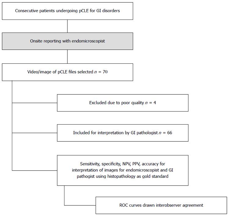
Figure 3 Flowchart of patient recruitment.
pCLE: Probe-based confocal laser endomicroscopy; PPV: Positive predictive value; NPV: Negative predictive value.
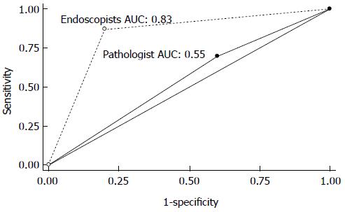
Figure 4 Receiver operating characteristic curves for endoscopists and pathologist for diagnosis of dysplastic/neoplastic lesions using probe-based confocal laser endomicroscopy.
- Citation: Peter S, Council L, Bang JY, Neumann H, Mönkemüller K, Varadarajulu S, Wilcox CM. Poor agreement between endoscopists and gastrointestinal pathologists for the interpretation of probe-based confocal laser endomicroscopy findings. World J Gastroenterol 2014; 20(47): 17993-18000
- URL: https://www.wjgnet.com/1007-9327/full/v20/i47/17993.htm
- DOI: https://dx.doi.org/10.3748/wjg.v20.i47.17993


