Copyright
©2014 Baishideng Publishing Group Inc.
World J Gastroenterol. Dec 14, 2014; 20(46): 17670-17673
Published online Dec 14, 2014. doi: 10.3748/wjg.v20.i46.17670
Published online Dec 14, 2014. doi: 10.3748/wjg.v20.i46.17670
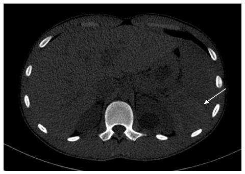
Figure 1 Computed tomography (white arrow) shows a hematoma at the upper pole of spleen.
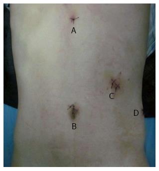
Figure 2 Size and distribution of trocars.
A: Subxiphoid, 5-mm; B: Upper umbilicus, 10-mm; C: Left mid clavicular line, 12-mm; D: Left axillary line, 5-mm.
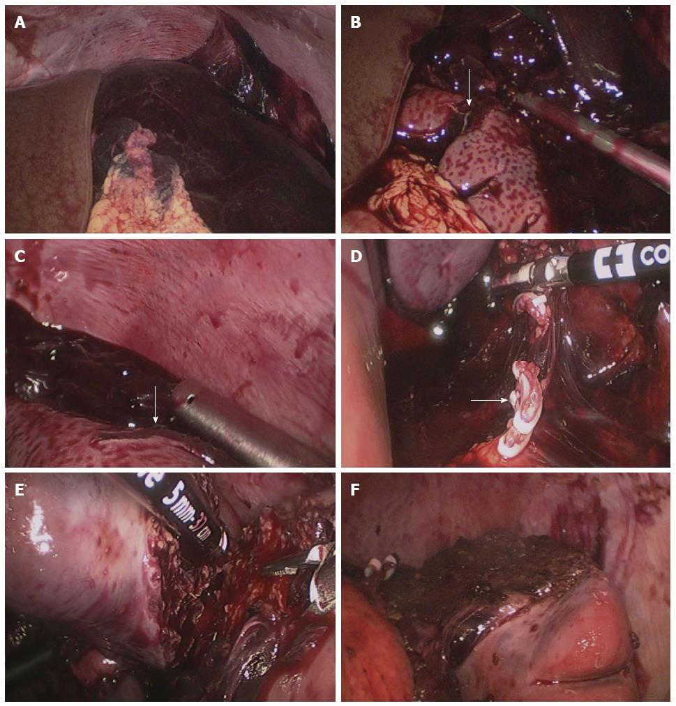
Figure 3 Intra-operative images.
A: A large hemoperitoneum surrounding the spleen; B: The anterior rupture of the spleen (white arrow); C: The posterior rupture of the spleen (white arrow); D: The branches of splenic artery were dissected; E: The parenchyma of spleen was dissected using a LigaSure vessel sealing system; F: The cut edge of the splenic stump.
- Citation: Cai YQ, Li CL, Zhang H, Wang X, Peng B. Emergency laparoscopic partial splenectomy for ruptured spleen: A case report. World J Gastroenterol 2014; 20(46): 17670-17673
- URL: https://www.wjgnet.com/1007-9327/full/v20/i46/17670.htm
- DOI: https://dx.doi.org/10.3748/wjg.v20.i46.17670


