Copyright
©2014 Baishideng Publishing Group Inc.
World J Gastroenterol. Dec 7, 2014; 20(45): 17244-17246
Published online Dec 7, 2014. doi: 10.3748/wjg.v20.i45.17244
Published online Dec 7, 2014. doi: 10.3748/wjg.v20.i45.17244
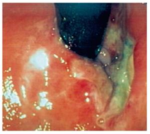
Figure 1 Ulcer located at the anterior part of the low rectum in retroflex examination with rectosigmoidoscopy.
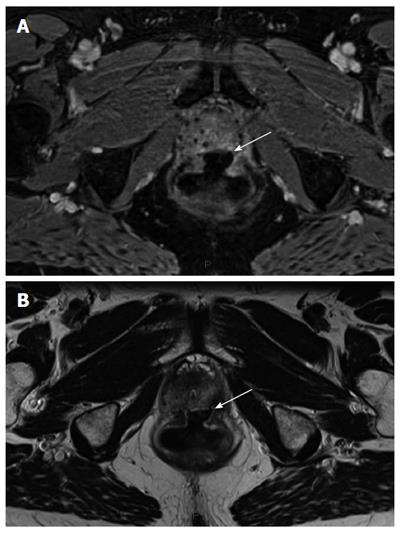
Figure 2 Large ulceration (indicated by white arrow) with regular edges sitting without local infectious complication or digestive fistula on pelvic magnetic resonance imaging performed 1 mo after endoscopic argon plasma coagulationprocedure.
A: Axial T1-weighted section after injection of gadolinium chelate; B: Axial T2-weighted section.
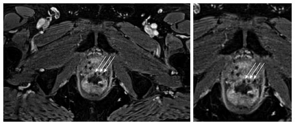
Figure 3 Presence of 125Iodine seed implants in an abnormal position.
Presence of 125Iodine seed implants in an abnormal position (arrows), sitting in the outer part of the anterior rectal wall instead of the large ulceration on pelvic magnetic resonance imaging performed 6 mo before endoscopic argon plasma coagulation procedure with axial T1-weighted section after injection of gadolinium chelate.
- Citation: Koessler T, Servois V, Mariani P, Aubert E, Cacheux W. Rectal ulcer: Due to ketoprofen, argon plasma coagulation and prostatic brachytherapy. World J Gastroenterol 2014; 20(45): 17244-17246
- URL: https://www.wjgnet.com/1007-9327/full/v20/i45/17244.htm
- DOI: https://dx.doi.org/10.3748/wjg.v20.i45.17244


