Copyright
©2014 Baishideng Publishing Group Inc.
World J Gastroenterol. Dec 7, 2014; 20(45): 17115-17119
Published online Dec 7, 2014. doi: 10.3748/wjg.v20.i45.17115
Published online Dec 7, 2014. doi: 10.3748/wjg.v20.i45.17115
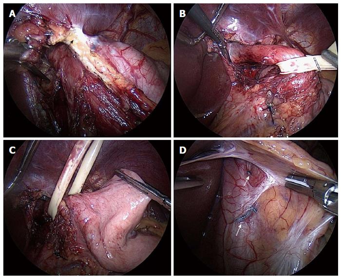
Figure 1 Intraoperative view.
A: The mobilization (arrow) of the prior transoral incisionless fundoplication; B: The mobilization of the esophagus with at least 3 cm within the abdominal cavity; C: The mobilization of the gastric fundus for a 360º Nissen fundoplication; D: The prior transoral incisionless fundoplication fasteners visible under the serosa of the stomach attached to the esophageal wall (arrows).
- Citation: Ashfaq A, Rhee HK, Harold KL. Revision of failed transoral incisionless fundoplication by subsequent laparoscopic Nissen fundoplication. World J Gastroenterol 2014; 20(45): 17115-17119
- URL: https://www.wjgnet.com/1007-9327/full/v20/i45/17115.htm
- DOI: https://dx.doi.org/10.3748/wjg.v20.i45.17115


