Copyright
©2014 Baishideng Publishing Group Inc.
World J Gastroenterol. Nov 28, 2014; 20(44): 16544-16549
Published online Nov 28, 2014. doi: 10.3748/wjg.v20.i44.16544
Published online Nov 28, 2014. doi: 10.3748/wjg.v20.i44.16544
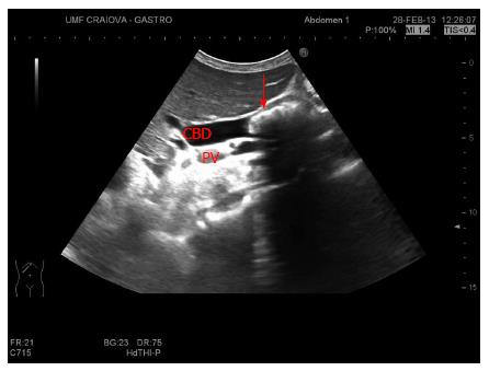
Figure 1 Large, conglomerated stones into a dilated common bile duct (over 12 mm).
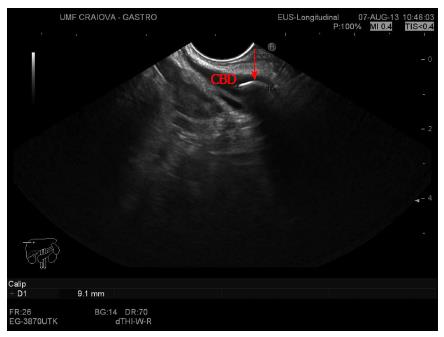
Figure 2 A 9 mm stone, within a slightly dilated, elongated common bile duct.
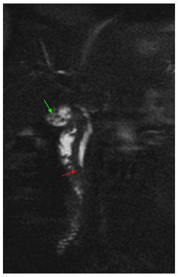
Figure 3 Multiple gallstones in T2 hyposignal less than 5 mm diameter (green arrow), diameter of the common bile duct 6 mm, with a 4 mm migrated stone (red arrow).
- Citation: Surlin V, Săftoiu A, Dumitrescu D. Imaging tests for accurate diagnosis of acute biliary pancreatitis. World J Gastroenterol 2014; 20(44): 16544-16549
- URL: https://www.wjgnet.com/1007-9327/full/v20/i44/16544.htm
- DOI: https://dx.doi.org/10.3748/wjg.v20.i44.16544


