Copyright
©2014 Baishideng Publishing Group Inc.
World J Gastroenterol. Nov 28, 2014; 20(44): 16474-16479
Published online Nov 28, 2014. doi: 10.3748/wjg.v20.i44.16474
Published online Nov 28, 2014. doi: 10.3748/wjg.v20.i44.16474
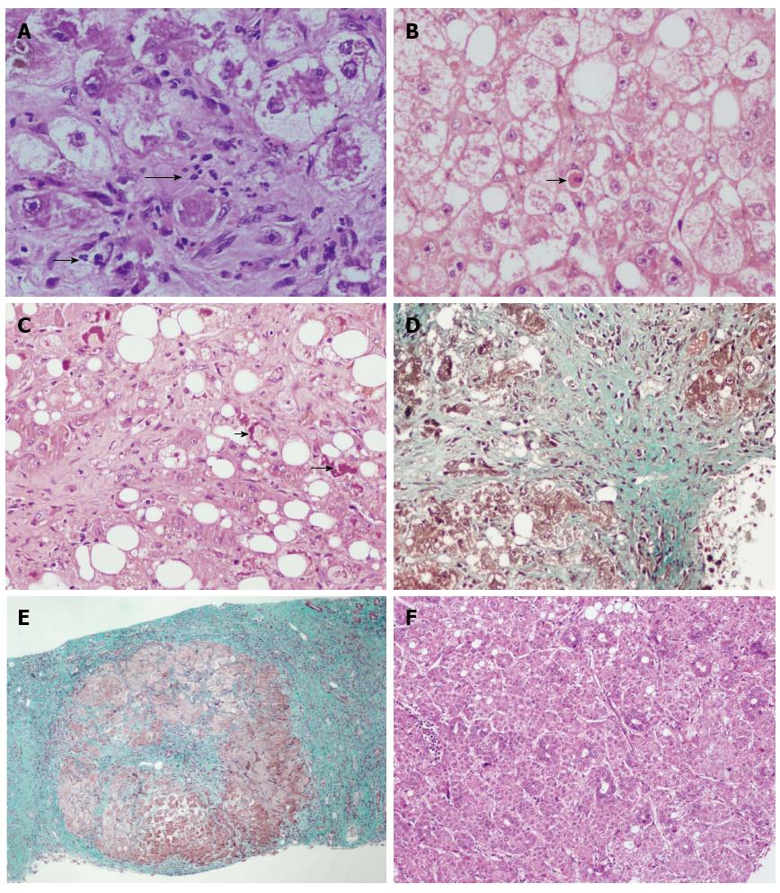
Figure 1 Histological image.
A: Neutrophilic inflammation (arrow) and ballooning of hepatocytes in alcoholic liver disease (ALD) (HE staining, × 400); B: Apoptotic cell death (arrow) in ALD (HE staining, × 400); C: Prominent Mallory hyaline (arrow) in ALD (HE staining, × 400); D: Sclerosing hyaline necrosis (masson trichrome staining, × 200); E: Cirrhosis with associated pericellular fibrosis also seen in ALD (masson trichrome staining, × 40); F: Hepatocellular carcinoma with focal steatosis in a case of known ALD (HE staining, × 100).
- Citation: Sakhuja P. Pathology of alcoholic liver disease, can it be differentiated from nonalcoholic steatohepatitis? World J Gastroenterol 2014; 20(44): 16474-16479
- URL: https://www.wjgnet.com/1007-9327/full/v20/i44/16474.htm
- DOI: https://dx.doi.org/10.3748/wjg.v20.i44.16474


