Copyright
©2014 Baishideng Publishing Group Inc.
World J Gastroenterol. Nov 21, 2014; 20(43): 16355-16358
Published online Nov 21, 2014. doi: 10.3748/wjg.v20.i43.16355
Published online Nov 21, 2014. doi: 10.3748/wjg.v20.i43.16355
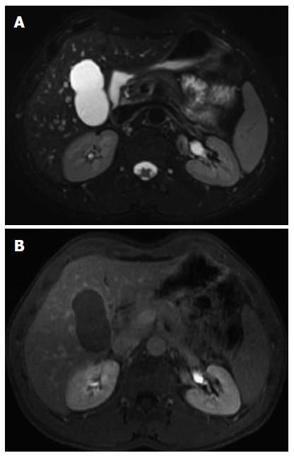
Figure 1 Magnetic resonance imaging.
A: With contrast; B: Without contrast. Magnetic resonance imaging demonstrates a 6.2 cm multilocular cystic lesion in segment 4. The cyst content was heterogeneous and avascular and included proteinaceous material. No wall enhancement was detected.
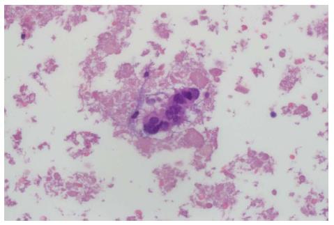
Figure 2 A computed tomography-guided diagnostic fine-needle aspiration revealed ciliated epithelial cells with no atypia.
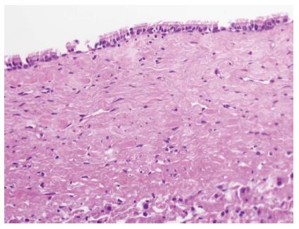
Figure 3 Wall of the cyst.
Histologically, the wall of the cyst showed characteristic pseudopapillae lined by ciliated stratified columnar epithelium, loos subepithelial connective tissue, underlying smooth muscle and an outer fibrous layer and no atypia, compatible with diagnosis of a ciliated hepatic foregut cyst.
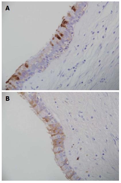
Figure 4 Immunohistochemistry.
A: Carbohydrate antigen (CA) 19-9: Some cells, including ciliated ones show apical staining for CA19-9; B: Carcinoembryonic antigen (CEA): Few cells show weak positivity for CEA.
- Citation: Ben Ari Z, Cohen-Ezra O, Weidenfeld J, Bradichevsky T, Weitzman E, Rimon U, Inbar Y, Amitai M, Bar-Zachai B, Eshkenazy R, Ariche A, Azoulay D. Ciliated hepatic foregut cyst with high intra-cystic carbohydrate antigen 19-9 level. World J Gastroenterol 2014; 20(43): 16355-16358
- URL: https://www.wjgnet.com/1007-9327/full/v20/i43/16355.htm
- DOI: https://dx.doi.org/10.3748/wjg.v20.i43.16355


