Copyright
©2014 Baishideng Publishing Group Inc.
World J Gastroenterol. Nov 14, 2014; 20(42): 15937-15940
Published online Nov 14, 2014. doi: 10.3748/wjg.v20.i42.15937
Published online Nov 14, 2014. doi: 10.3748/wjg.v20.i42.15937
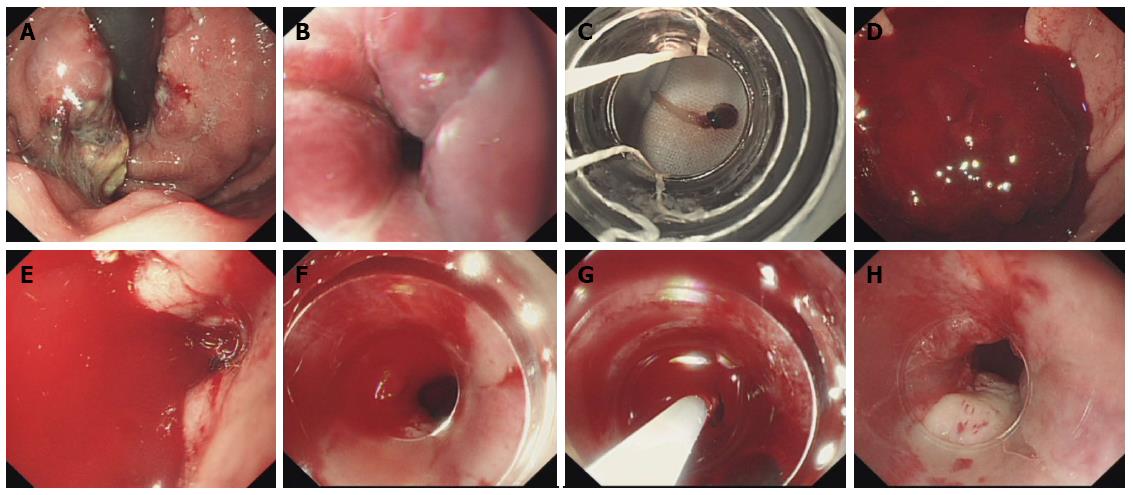
Figure 1 Esophagogastroduodenoscopy revealed incomplete tissue glue extrusion and massive bleeding.
A: Tissue glue extrusion was shown via endoscopy; B: Straight esophageal varices with red color signs were observed in the cardiac area; C: A piece of tadpole-shaped, semi-transparent, semi-soft tissue glue partially covered by a small piece of mucosa and blood vessel wall adhering to the transparent cap was released from the cap; D: Massive bleeding was revealed; E: The endoscopic vision was poor without the aid of the transparent cap; F: Massive, spurting bleeding was observed with the aid of the transparent cap; G: A precise intravenous injection was achieved with the aid of the transparent cap; H: The bleeding ceased.
- Citation: Wei XQ, Gu HY, Wu ZE, Miao HB, Wang PQ, Wen ZF, Wu B. Endoscopic variceal ligation caused massive bleeding due to laceration of an esophageal varicose vein with tissue glue emboli. World J Gastroenterol 2014; 20(42): 15937-15940
- URL: https://www.wjgnet.com/1007-9327/full/v20/i42/15937.htm
- DOI: https://dx.doi.org/10.3748/wjg.v20.i42.15937


