Copyright
©2014 Baishideng Publishing Group Inc.
World J Gastroenterol. Nov 14, 2014; 20(42): 15920-15924
Published online Nov 14, 2014. doi: 10.3748/wjg.v20.i42.15920
Published online Nov 14, 2014. doi: 10.3748/wjg.v20.i42.15920
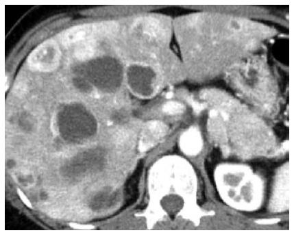
Figure 1 Abdominal enhanced computed tomography showed a contrast-enhanced tumor lesions accompanied by cystic changes in the liver and the pancreatic tail.
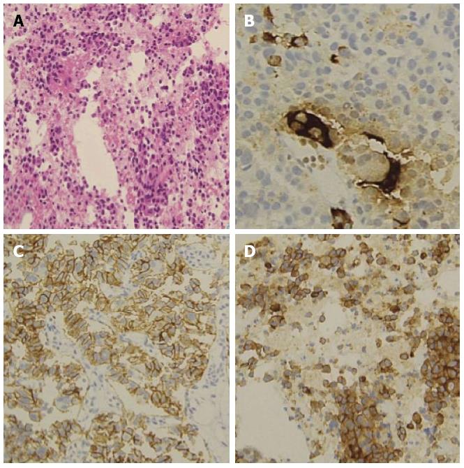
Figure 2 Endoscopic ultrasound-fine needle aspiration revealed a neuroendocrine tumor.
A: Hematoxylin and eosin; B: Chromogranin A; C: CD56; D: Synaptophisin.
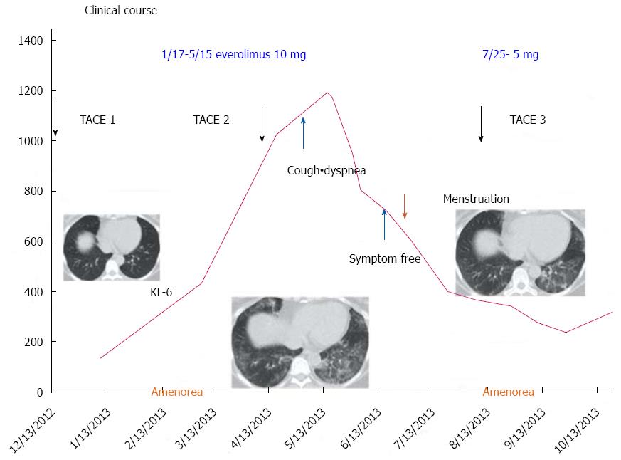
Figure 3 Clinical course.
TACE: Transcatheter arterial chemoembolization.
- Citation: Kawaguchi Y, Maruno A, Kawashima Y, Ito H, Ogawa M, Mine T. Amenorrhea as a rare drug-related adverse event associated with everolimus for pancreatic neuroendocrine tumors. World J Gastroenterol 2014; 20(42): 15920-15924
- URL: https://www.wjgnet.com/1007-9327/full/v20/i42/15920.htm
- DOI: https://dx.doi.org/10.3748/wjg.v20.i42.15920


