Copyright
©2014 Baishideng Publishing Group Inc.
World J Gastroenterol. Nov 14, 2014; 20(42): 15797-15804
Published online Nov 14, 2014. doi: 10.3748/wjg.v20.i42.15797
Published online Nov 14, 2014. doi: 10.3748/wjg.v20.i42.15797
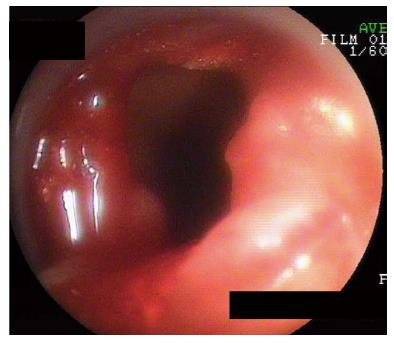
Figure 1 Endoscope-related perforation.
A perforation of the long limb with Billroth II anastomosis that occurred during double balloon enteroscope insertion and was detected immediately by endoscopic view (Case 1).
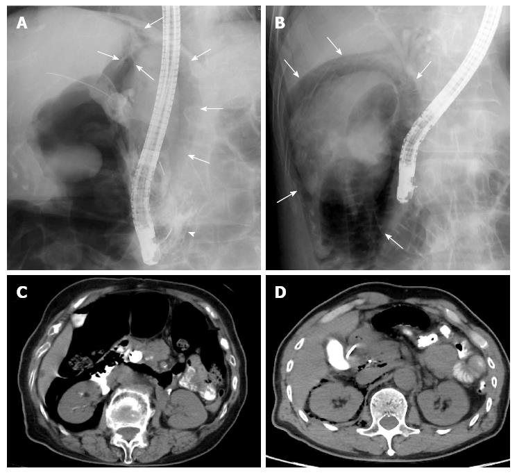
Figure 2 Endoscopic sphincterotomy-related perforations.
A: Retroperitoneal free air (arrows) and contrast media leakage (arrowhead) were observed by fluoroscopy (Case 2); B: Retroperitoneal free air (arrows) was observed by fluoroscopy (Case 5); C: Retroperitoneal free air and contrast media leakage was detected by a computed tomography (CT) scan performed immediately after surgery (Case 2); D: Retroperitoneal free air was detected by CT scan performed immediately after surgery (Case 5).
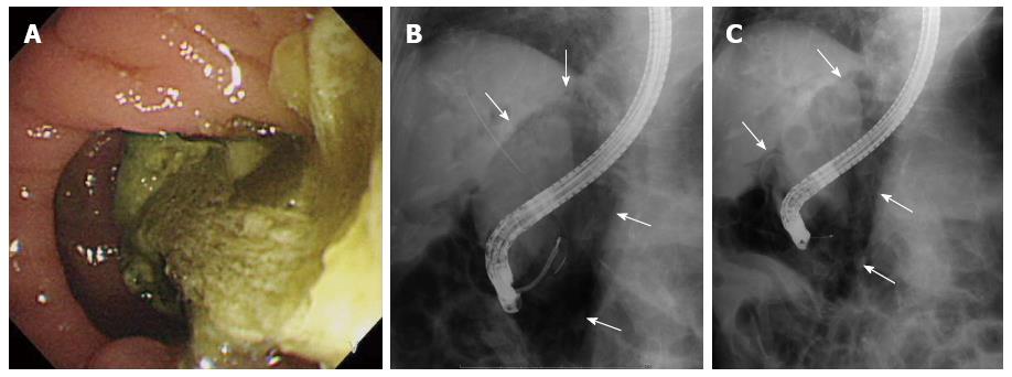
Figure 3 Endoscopic instrument-related perforation.
A: A food residue of substantial volume was observed in the periampullary diverticula prior to the endoscopic retrograde cholangiopancreatography (ERCP) procedure (Case 3); B: Retroperitoneal free air (arrows) was detected by fluoroscopy after endoscopic sphincterotomy (EST) was performed during the ERCP procedure; C: The free air (arrows) was present before EST was performed.
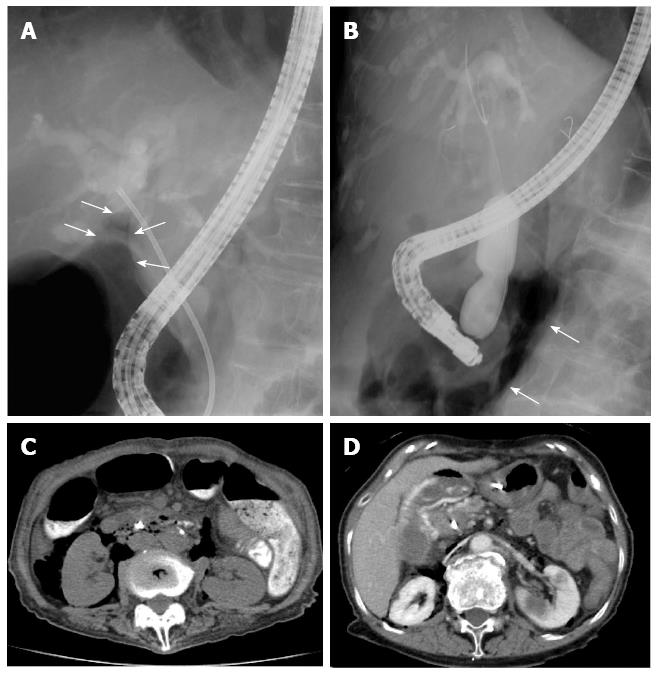
Figure 4 Endoscopic papillary balloon dilation-related perforations.
A: Retroperitoneal free air (arrows) was detected by fluoroscopy (Case 4); B: Retroperitoneal free air (arrows) was detected by fluoroscope during balloon dilation (Case 6); C: Retroperitoneal free air was detected by computed tomography (CT) scan performed immediately after surgery (Case 4); D: Retroperitoneal free air was detected by CT scan performed immediately after surgery (Case 6).
- Citation: Motomura Y, Akahoshi K, Gibo J, Kanayama K, Fukuda S, Hamada S, Otsuka Y, Kubokawa M, Kajiyama K, Nakamura K. Immediate detection of endoscopic retrograde cholangiopancreatography-related periampullary perforation: Fluoroscopy or endoscopy? World J Gastroenterol 2014; 20(42): 15797-15804
- URL: https://www.wjgnet.com/1007-9327/full/v20/i42/15797.htm
- DOI: https://dx.doi.org/10.3748/wjg.v20.i42.15797


