Copyright
©2014 Baishideng Publishing Group Inc.
World J Gastroenterol. Nov 14, 2014; 20(42): 15771-15779
Published online Nov 14, 2014. doi: 10.3748/wjg.v20.i42.15771
Published online Nov 14, 2014. doi: 10.3748/wjg.v20.i42.15771
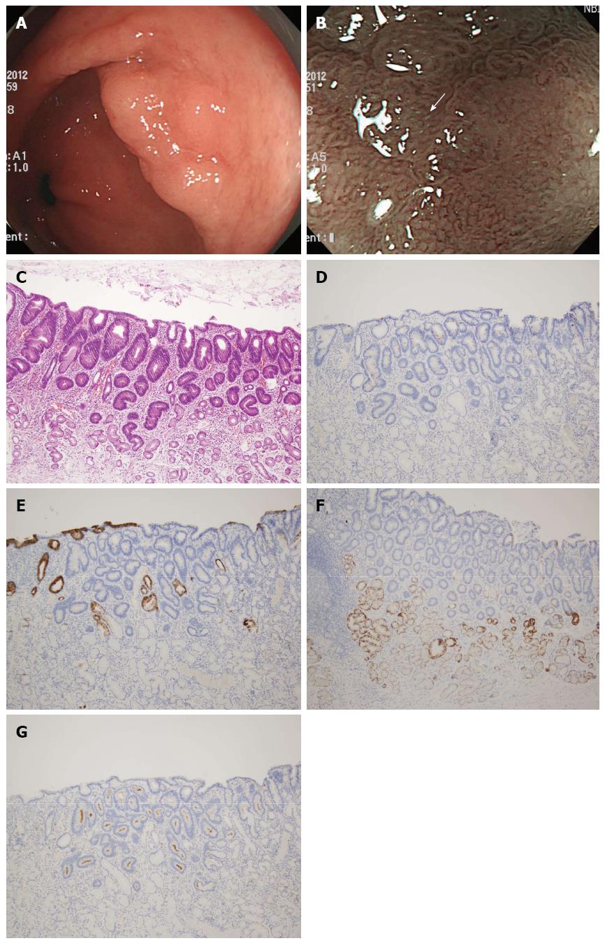
Figure 1 Endoscopic and histologic findings of adenomatous gastric epithelial dysplasia.
A: An elevated lesion with nodular changes is seen at the antrum; B: Magnifying endoscopy using narrow band imaging shows a mixed (tubular and round pit) microsurface and regular microvascular pattern. Light blue crests are also seen (arrow); C: Histologic examination shows that the dysplasia is composed of tubules lined by columnar cells with hyperchromatic, pencillate nuclei with pseudostratification (Haematoxylin-Eosin stain, × 10). The dysplastic epithelium is strongly positive for CD10 (G) and negative for MUC2 (D), MUC5AC (E) and MUC6 (F) (Immunohistochemistry, × 10).
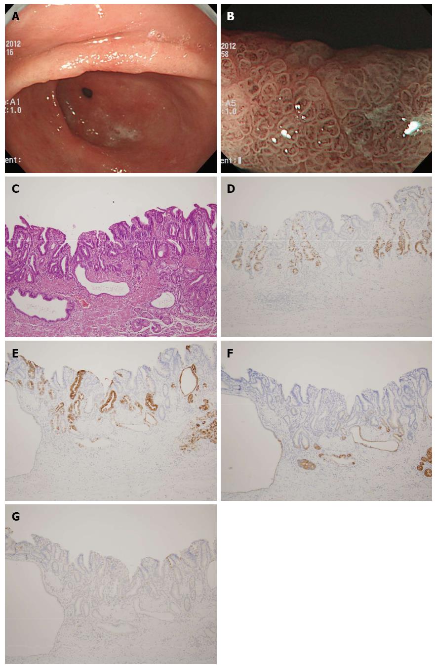
Figure 2 Endoscopic and histologic findings of foveolar gastric epithelial dysplasia.
A: A slightly depressed lesion is seen at the antrum; B: Magnifying endoscopy using narrow band imaging shows a papillary microsurface and slightly irregular microvascular pattern; C: Histologic examination shows that the dysplasia is composed of cuboidal to columnar cells with a pale cytoplasm and basally located ovoid nuclei. Hyperplasia of the foveolar region with irregular glandular branching and epithelial folding are also seen (Haematoxylin-Eosin stain, × 10). The dysplastic epithelium is strongly positive for MUC5AC (E) and negative for MUC2 (D), MUC6 (F) and CD10 (G) (Immunohistochemistry, × 10).
- Citation: Kang HM, Kim GH, Park DY, Cheong HR, Baek DH, Lee BE, Song GA. Magnifying endoscopy of gastric epithelial dysplasia based on the morphologic characteristics. World J Gastroenterol 2014; 20(42): 15771-15779
- URL: https://www.wjgnet.com/1007-9327/full/v20/i42/15771.htm
- DOI: https://dx.doi.org/10.3748/wjg.v20.i42.15771


