Copyright
©2014 Baishideng Publishing Group Inc.
World J Gastroenterol. Nov 14, 2014; 20(42): 15664-15673
Published online Nov 14, 2014. doi: 10.3748/wjg.v20.i42.15664
Published online Nov 14, 2014. doi: 10.3748/wjg.v20.i42.15664
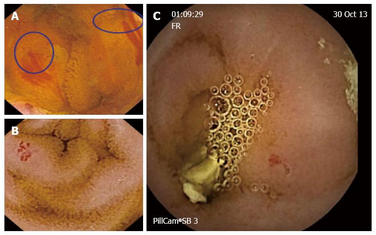
Figure 1 Active bleeding and angiodysplasias in patients with obscure gastrointestinal bleeding.
A: Active bleeding; B and C: Angiodysplasias in patients with obscure gastrointestinal bleeding.
Figure 2 Ulcers in the small bowel.
A: Ulcer with erythematous and edematous edges; B: Serpiginous ulceration and stenosis affecting the entire colon circumference.
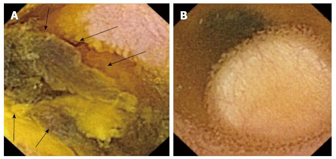
Figure 3 Tumors in the small bowel.
A: Dark tumor (arrows) confirming metastases from melanoma after surgery; B: Subepithelial white lesion compatible with lipoma.
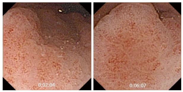
Figure 4 Enlarged gastric areas in a patient with Peutz Jeghers syndrome.
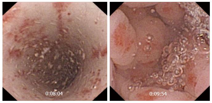
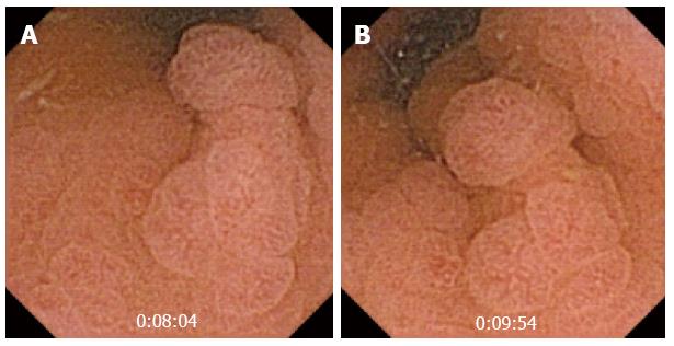
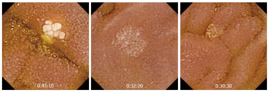
Figure 5 Small bowel lymphangiectasia.
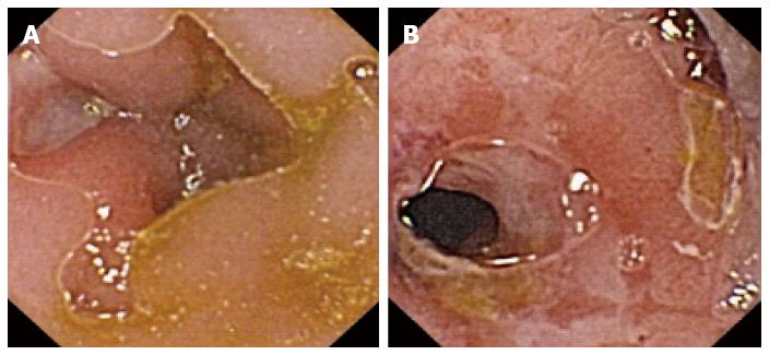
Figure 6 Erythematous lesions in the upper gastrointestinal tract.
A: Erythematous esophageal lesions in a patient with gastrointestinal amyloidosis; B: Gastric lesions in patients with obscure gastrointestinal bleeding.
Figure 7 Portal hypertension related gastropathy in a cirrhotic patient.
- Citation: Redondo-Cerezo E, Sánchez-Capilla AD, De La Torre-Rubio P, De Teresa J. Wireless capsule endoscopy: Perspectives beyond gastrointestinal bleeding. World J Gastroenterol 2014; 20(42): 15664-15673
- URL: https://www.wjgnet.com/1007-9327/full/v20/i42/15664.htm
- DOI: https://dx.doi.org/10.3748/wjg.v20.i42.15664


