Copyright
©2014 Baishideng Publishing Group Inc.
World J Gastroenterol. Nov 14, 2014; 20(42): 15539-15548
Published online Nov 14, 2014. doi: 10.3748/wjg.v20.i42.15539
Published online Nov 14, 2014. doi: 10.3748/wjg.v20.i42.15539
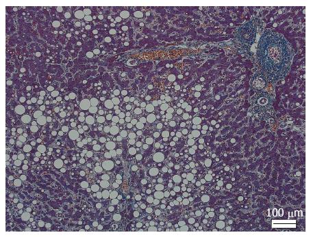
Figure 1 Steatosis in nonalcoholic fatty liver disease.
Macrovesicular steatosis, predominantly distributed in zone 3, is observed (Masson trichrome staining).
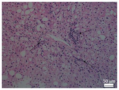
Figure 2 Lobular inflammation in nonalcoholic steatohepatitis.
Necroinflammatory foci (arrows) are scattered in the hepatic lobule (hematoxylin and eosin staining).
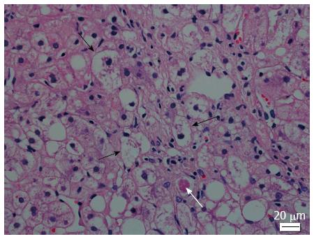
Figure 3 Ballooning and Mallory-Denk bodies in nonalcoholic steatohepatitis.
Ballooned hepatocytes are recognized as swollen hepatocytes with rarefied cytoplasm (black arrows). Mallory-Denk bodies are eosinophilic irregular-shaped aggregates in the cytoplasm of hepatocytes (white arrow) (hematoxylin and eosin staining).
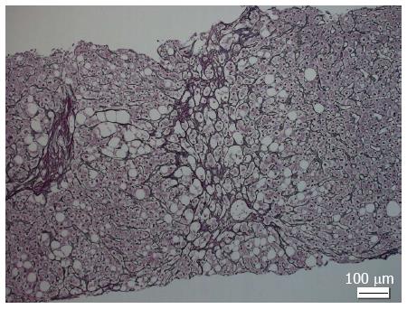
Figure 4 Fibrosis in nonalcoholic steatohepatitis.
The characteristic pattern of fibrosis in nonalcoholic steatohepatitis is perisinusoidal/pericellular (chicken wire) fibrosis, which usually begins in zone 3 (reticulin staining).
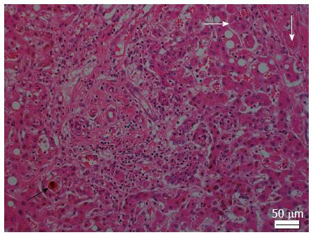
Figure 5 Histological appearance of alcoholic steatohepatitis.
Canalicular cholestasis (black arrow), Mallory-Denk bodies (white arrows), and acute inflammation in the portal tract are observed (hematoxylin and eosin staining).
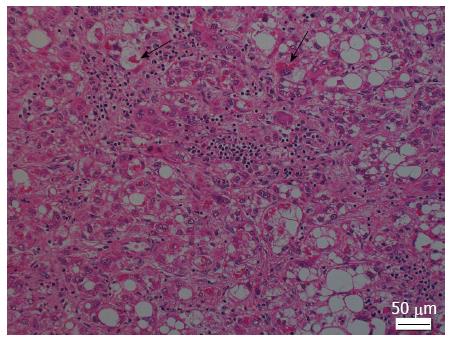
Figure 6 Histological appearance of steatohepatitic hepatocellular carcinoma.
Large droplet steatosis, ballooning of malignant hepatocytes, Mallory-Denk bodies (arrows), inflammation, and fibrosis are observed in tumor tissue (hematoxylin and eosin staining).
- Citation: Takahashi Y, Fukusato T. Histopathology of nonalcoholic fatty liver disease/nonalcoholic steatohepatitis. World J Gastroenterol 2014; 20(42): 15539-15548
- URL: https://www.wjgnet.com/1007-9327/full/v20/i42/15539.htm
- DOI: https://dx.doi.org/10.3748/wjg.v20.i42.15539


