Copyright
©2014 Baishideng Publishing Group Inc.
World J Gastroenterol. Nov 14, 2014; 20(42): 15499-15517
Published online Nov 14, 2014. doi: 10.3748/wjg.v20.i42.15499
Published online Nov 14, 2014. doi: 10.3748/wjg.v20.i42.15499
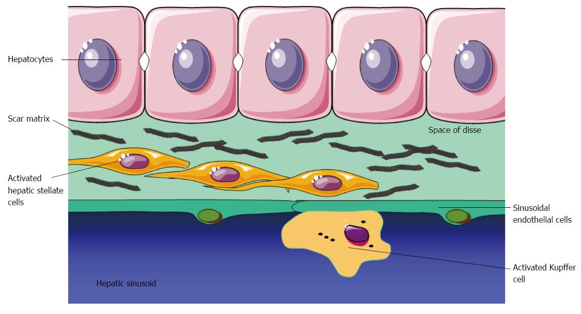
Figure 1 Fibrogenesis after liver injury.
Hepatic stellate cells are activated into myofibroblasts that deposit scar matrix in the Space of Disse.
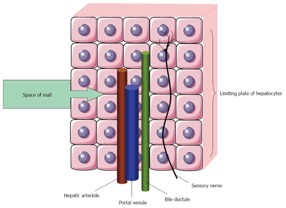
Figure 2 Space of Mall is a small space of fluid surrounding a hepatic arteriole, a portal venule and a bile ductule.
Adenosine is secreted into the space of Mall. A reduction in portal blood flow increases adenosine levels and leads to hepatic arteriolar vasodilataion and activation of sensory nerves after[54].
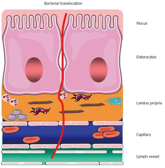
Figure 3 Illustration of bacterial translocation from the gastrointestinal lumen through the epithelial layers and capillaries to the lymphalic vessels.
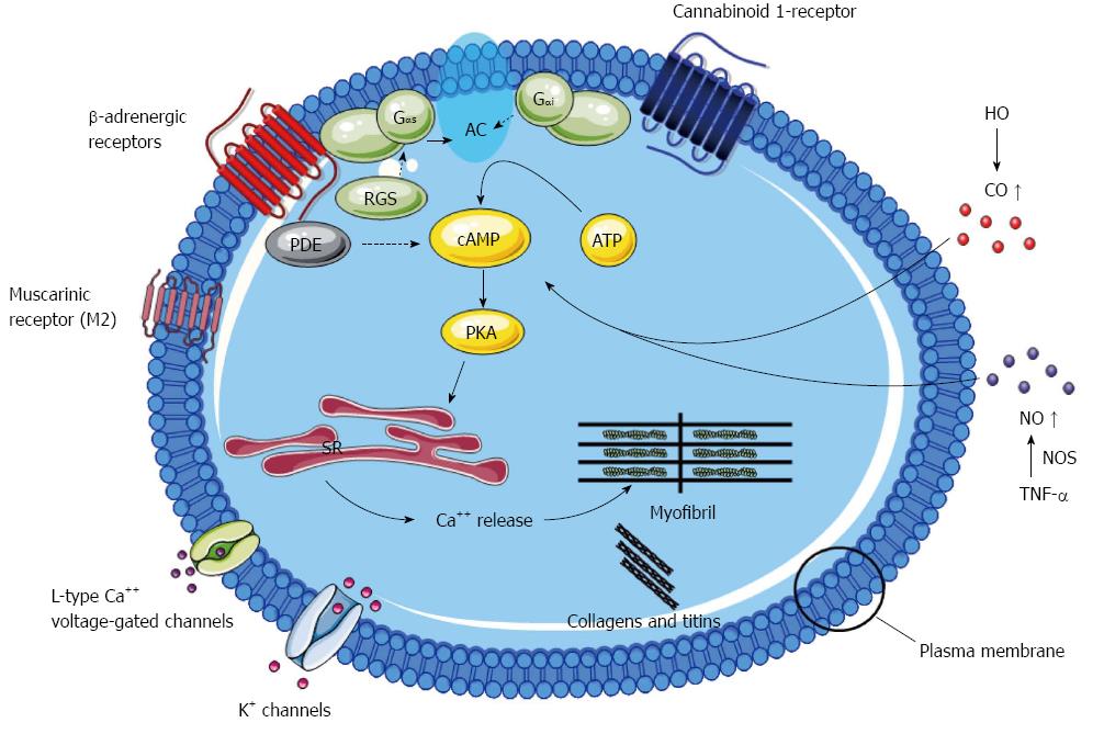
Figure 4 Mechanisms of cirrhotic cardiomyopathy.
The figure reviews the most important mechanisms involved in cirrhotic cardiomyopathy: Desensitisation and downregulation of β-adrenergic receptors with decreased content of G-protein (Gαi: inhibitory G protein; Gαs: stimulatory G protein) and following impaired intracellular signalling; alterations in particular in M2 muscarinic receptors; upregulation of cannabinoid 1-receptor stimulation; altered plasma membrane cholesterol/phospholipid ratio; increased inhibitory effects of haemooxygenase (HO), carbon monoxide (CO), nitric oxide (NO), and tumour necrosis factor-α (TNF-α); reduced density of potassium channels; changed function and fluxes through L-type calcium channels; altered ratio and function of collagens and titins. Many post-receptor effects are mediated by adenylcyclase (AC) inhibition or stimulation. PKA: Protein kinase A.
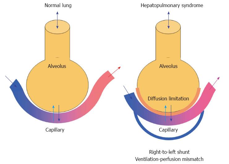
Figure 5 Gas exchange in the normal lung (left) and mechanism of hepatopulmonary syndrome (right).
The hepatopulmonary syndrome comprises an increased alveolar-arterial oxygen gradient owing to diffusion limitations and development of intrapulmonary right-to-left shunts leading to arterial hypoxaemia.
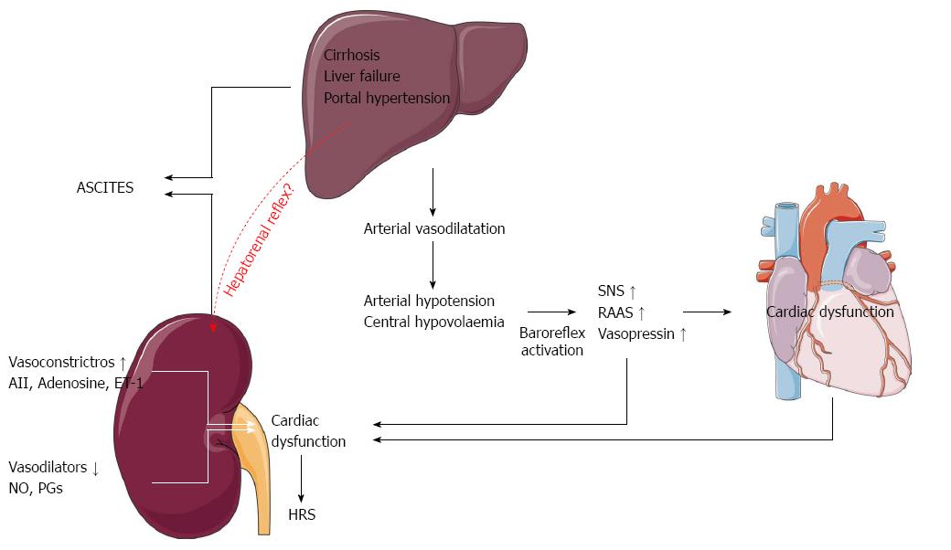
Figure 6 Pathophysiological mechanisms in the development of ascites and the hepatorenal syndrome.
SNS: Sympathetic nervous system; RAAS: The renin-angiotensin-aldosterone system; AII: Angiotensin II; ET-1: Endothelin-1; NO: Nitric oxide; PGs: Prostaglandins.
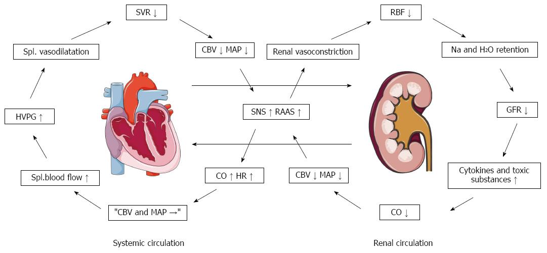
Figure 7 Pathophysiological proposal for the background of a cardiorenal syndrome in cirrhosis[194].
CBV: Central blood volume; CO: Cardiac output; GFR: Glomerular filtration rate; HR: Heart rate; HVPG: Hepatic venous pressure gradient; MAP: Mean arterial pressure; RAAS: Renin-angiotensin-aldosterone system; RBF: Renal blood flow; SNS: Sympathetic nervous system; SVR: Systemic vascular resistance.
- Citation: Møller S, Henriksen JH, Bendtsen F. Extrahepatic complications to cirrhosis and portal hypertension: Haemodynamic and homeostatic aspects. World J Gastroenterol 2014; 20(42): 15499-15517
- URL: https://www.wjgnet.com/1007-9327/full/v20/i42/15499.htm
- DOI: https://dx.doi.org/10.3748/wjg.v20.i42.15499


