Copyright
©2014 Baishideng Publishing Group Inc.
World J Gastroenterol. Nov 7, 2014; 20(41): 15454-15461
Published online Nov 7, 2014. doi: 10.3748/wjg.v20.i41.15454
Published online Nov 7, 2014. doi: 10.3748/wjg.v20.i41.15454
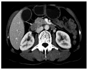
Figure 1 Preoperative radiological findings of a duodenal lesion.
Computed tomography scan revealed that a 3.0 cm duodenal mass in the medial wall of the subpapillary duodenum (white arrow).
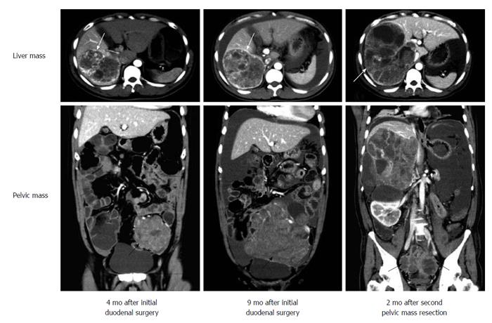
Figure 2 Postoperative radiological findings in follow-up periods.
Computed tomography (CT) scans at 4 mo and 9 mo after initial duodenal surgery showed distant metastatic lesions located in the liver (white arrows) and pelvic cavity (black arrows). The mass in the pelvic cavity became larger and pressed the surrounding tissues. Two months after second pelvic surgery, CT scans showed that the mass re-grew in the pelvic cavity (black arrows) and the liver mass became larger (white arrows).
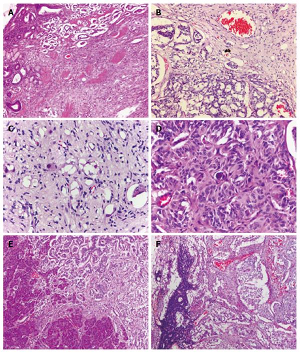
Figure 3 Photomicrographs of the duodenal lesion.
A: A low-power image of the duodenal lesion showed a mass occupying the submucosa and extending into muscularis propria with an infiltrative margin; B: At high-power fields, the lesion was composed of epithelioid cells arranged in a nested fashion, slender spindle-shaped cells and occasional ganglion-like cells; C, D: Occasional psammoma bodies could also be found in the lesion; The duodenal lesion exhibited a penetrative growth pattern with pancreatic infiltration (E) and regional lymph node metastasis (F). All three components of the tumor was found in effaced nodal architecture (A: HE staining with original magnification × 40; B-D: HE staining with original magnification × 400; E, F: HE staining with original magnification × 200). HE: Hematoxylin and eosin.
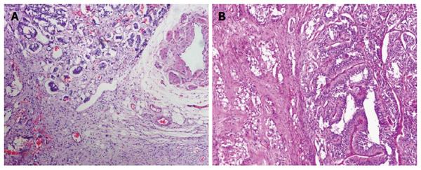
Figure 4 Photomicrographs of the pelvic cavity lesion.
A: At low-power fields, the lesion appeared to have an infiltrative margin without necrosis, hemorrhage or invasion of blood vessels; B: At high-power fields, the lesion exhibited an admixture of three types of tumor cells, and epithelioid cells were observed to be arranged in pseudoglandular structures (A: HE staining with original magnification × 100; B: HE staining with original magnification × 400). HE: Hematoxylin and eosin.
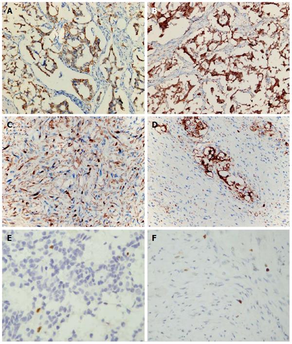
Figure 5 Immunohistochemical analysis of the lesions in the duodenum, lymph nodes and pelvic cavity.
The epithelioid cells were positive for cytokeratin (AE1/AE3) (A) and chromogranin A (B). The spindle-shaped cells were diffusely positive for S-100 protein (C), and ganglion-like cells were positive for synaptophysin (D). Ki-67 index was low in either epithelioid cell areas (E) or spindle-shaped cell areas (F). A-D: Immunohistochemical staining with original magnification × 400.
- Citation: Li B, Li Y, Tian XY, Luo BN, Li Z. Malignant gangliocytic paraganglioma of the duodenum with distant metastases and a lethal course. World J Gastroenterol 2014; 20(41): 15454-15461
- URL: https://www.wjgnet.com/1007-9327/full/v20/i41/15454.htm
- DOI: https://dx.doi.org/10.3748/wjg.v20.i41.15454


