Copyright
©2014 Baishideng Publishing Group Inc.
World J Gastroenterol. Nov 7, 2014; 20(41): 15448-15453
Published online Nov 7, 2014. doi: 10.3748/wjg.v20.i41.15448
Published online Nov 7, 2014. doi: 10.3748/wjg.v20.i41.15448
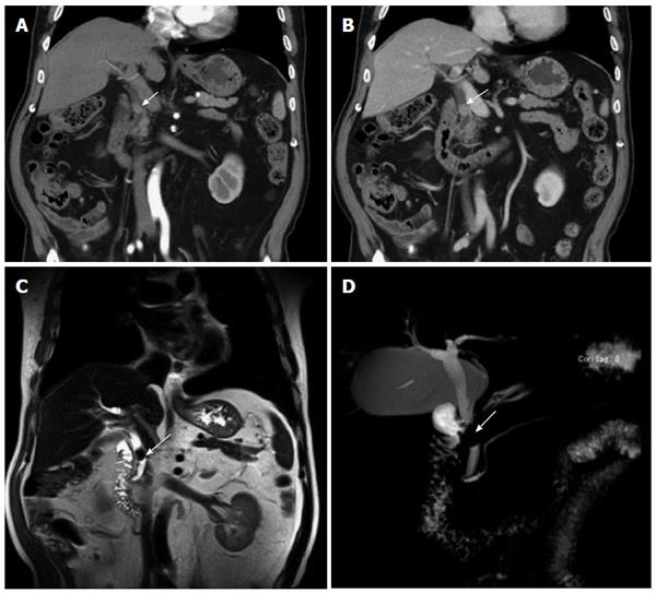
Figure 1 Preoperative images showing the tumor (arrows).
A: Non-enhanced upper-abdominal coronal computed tomography demonstrating a 1.2-cm mass in the middle of the common bile duct; B: Intravenous administration of contrast medium showing mild enhancement of the tumor; C: T2–weighted magnetic resonance image showed that the tumor had a relatively well-defined border; D: Magnetic resonance cholangiopancreatography image revealed stenosis of the common bile duct.
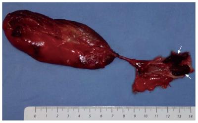
Figure 2 Resected specimen revealed a polypoid lesion (arrows) in the middle of the common bile duct.
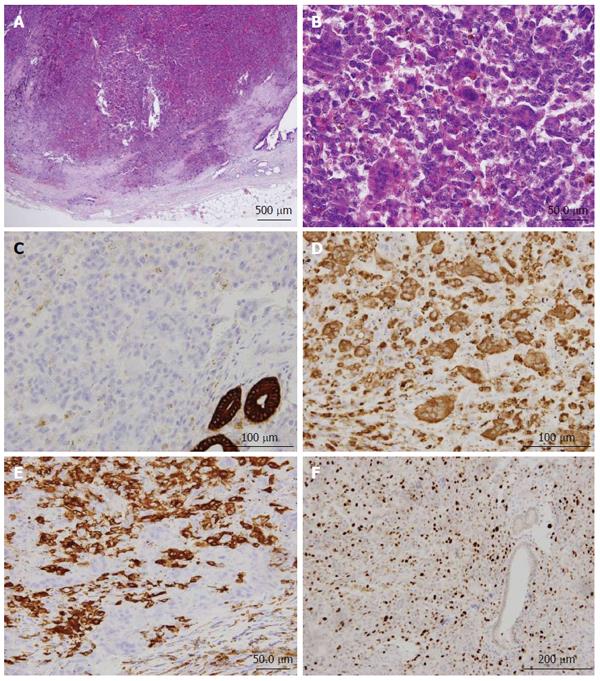
Figure 3 Photomicrographs showing the general appearance of the tumor.
A: Hematoxylin and eosin (HE) staining. A vague nodular pattern in the tumor and the normal surface epithelium of the common bile duct are observable. Note the infiltration into the wall; B: Mononuclear cells were mixed with multinucleated osteoclast-like giant cells. There was no cytologic atypia or mitotic figures, red-cell extravasation was present; HE staining; C: Tumor cells showing negative staining for CK19 compared with the remaining normal bile duct; D: Mononuclear cells and multinucleated giant cells showing strong cytoplasmic immunoreactivity for CD68; E: Granular cytoplasmic immunoreactivity for CD163 was restricted to the mononuclear cells; F: The mononuclear cells had an average Ki67 index of 10%-20%.
- Citation: Wang DD, Zheng YM, Teng LH, Sun YN, Gao W, Wang LM, Wang YH, Li F, Lu DH. Benign giant-cell tumor of the common bile duct: A case report. World J Gastroenterol 2014; 20(41): 15448-15453
- URL: https://www.wjgnet.com/1007-9327/full/v20/i41/15448.htm
- DOI: https://dx.doi.org/10.3748/wjg.v20.i41.15448


