Copyright
©2014 Baishideng Publishing Group Inc.
World J Gastroenterol. Nov 7, 2014; 20(41): 15216-15232
Published online Nov 7, 2014. doi: 10.3748/wjg.v20.i41.15216
Published online Nov 7, 2014. doi: 10.3748/wjg.v20.i41.15216
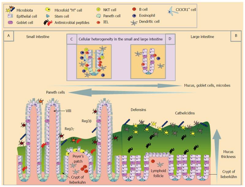
Figure 1 Schematic representation of the structural and cellular heterogeneity within the small and large intestine during homeostatic conditions.
The small (A and C) and large (B and D) intestine differ greatly in their structure and cellular composition. The small intestinal epithelium is folded to create crypts of Lieberkühn, has finger-like projections called villi and epithelial cell possess striated microvilli (A) whereas the large intestine (B) has crypts and lacks villi. The small intestine has Peyer’s patches, the main site of Microfold or “M” cells that transcytose luminal particulate antigens that are rare in the larger intestine. Both the small and large intestine are covered by mucus, which is much thicker in the large intestine (B) than in the small intestine (A). The large intestine houses the largest and most diverse microbial populations of the two sites (B). Anti-microbial peptides are also differentially expressed along the gastrointestinal tract. The distribution of immune cells also differ in the small and large intestine (inset C and D) with Natural killer T cells, Intraepithelial lymphocytes (IEL), eosinophils and dendritic cells found in the highest numbers in the small intestinal epithelium under steady state conditions. Cell populations are defined in the key.
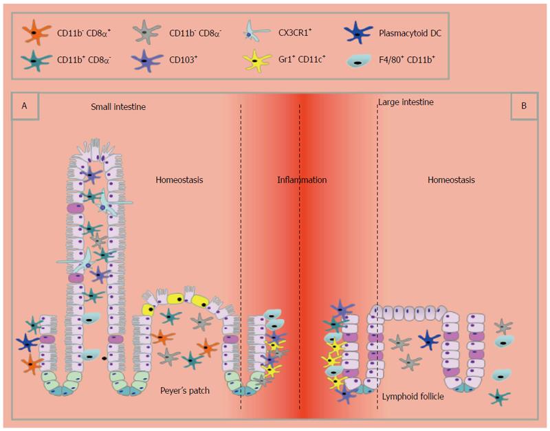
Figure 2 Schematic representation of the distribution of dendritic cell and macrophage subset in the small and large intestine under homeostatic conditions and upon inflammation.
In homeostasis dendritic cells (DCs) and macrophages are found in close contact with the epithelium in the small intestine (A) whereas in the large intestine they are rare and in the LP (B) and are recruited in response to inflammatory stimuli. Within the small intestine lymphoid resident DC (CD11chigh, MHCIIhigh CD64-) can be grouped into CD8α+, CD11b+ and CD8α-CD11b- DCs, which are located in Peyer’s patches. In the lamina propria (LP) of the small intestine CD11b+CD8α- DCs predominate. CD8α-CD11b- DCs have been identified in regions of the large intestine, including the ILFs, but CD11b-CD8α+ DCs are rarely found during homeostasis. Plasmacytoid B220+ CD11cintermediate DC are present in very low numbers in the LP of the small intestine and ILFs of the large intestine. In the small intestine CX3CR1+ cells can produce transepithelial dendrites that extend through the epithelium to directly sample luminal contents. This has not been demonstrated in the large intestine during homeostasis. In addition to CX3CR1, macrophages can be further defined by expression of CD11b, F4/80 and CD64. Upon inflammation, Gr1+ monocyte-derived inflammatory DCs accumulate in both the small and large intestine.
- Citation: Bowcutt R, Forman R, Glymenaki M, Carding SR, Else KJ, Cruickshank SM. Heterogeneity across the murine small and large intestine. World J Gastroenterol 2014; 20(41): 15216-15232
- URL: https://www.wjgnet.com/1007-9327/full/v20/i41/15216.htm
- DOI: https://dx.doi.org/10.3748/wjg.v20.i41.15216


