Copyright
©2014 Baishideng Publishing Group Inc.
World J Gastroenterol. Nov 7, 2014; 20(41): 15098-15109
Published online Nov 7, 2014. doi: 10.3748/wjg.v20.i41.15098
Published online Nov 7, 2014. doi: 10.3748/wjg.v20.i41.15098
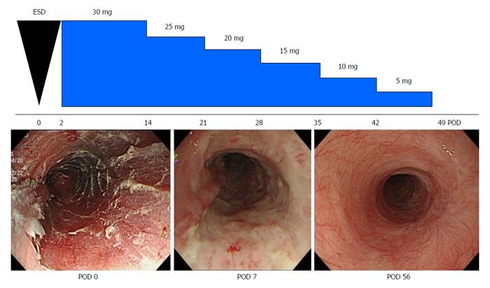
Figure 1 Systemic steroid therapy for the prevention of esophageal strictures after endoscopic submucosal dissection[83].
A 72-year-old man underwent esophageal endoscopic submucosal dissection (ESD) for early squamous cell carcinoma. Systemic steroid therapy with prednisolone was started at postoperative day 2. At postoperative day 51 following esophageal ESD, the ulcer surface was covered with a regenerative mucosa, and no esophageal stricture was present. Figures are reproduced courtesy of Shindan to Chiryo Sha, Inc. POD: Postoperative day.
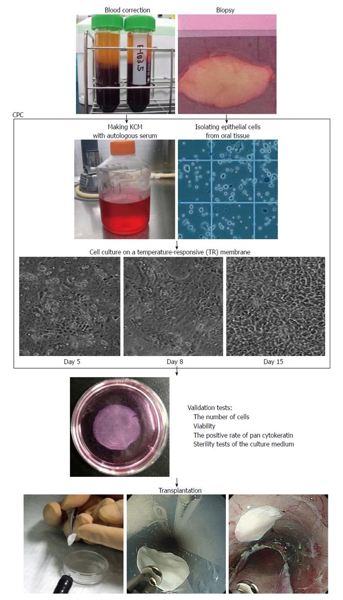
Figure 2 Protocol for the fabrication of autologous oral epithelial cell sheets for clinical application.
Autologous oral epithelial cell sheets are fabricated using autologous serum and oral mucosa in the cell processing center (CPC). Isolated oral epithelial cells are cultured on a temperature-responsive membrane for 16 d. The fabricated oral epithelial cell sheets are then transplanted endoscopically after passing several validation tests. KCM: Keratinocyte culture medium.
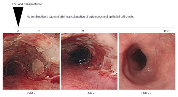
Figure 3 Transplantation of autologous oral epithelial cell sheets for the prevention of esophageal strictures after endoscopic submucosal dissection[65].
A 55-year-old man underwent esophageal endoscopic submucosal dissection (ESD) for early squamous cell carcinoma. Seven fabricated autologous oral epithelial cell sheets were transplanted immediately after esophageal ESD. At postoperative day 21, the ulcer surface after esophageal ESD was covered with a regenerative mucosa, and no esophageal stricture was present. Figures are reproduced courtesy of Elsevier. POD: Postoperative day.
- Citation: Kobayashi S, Kanai N, Ohki T, Takagi R, Yamaguchi N, Isomoto H, Kasai Y, Hosoi T, Nakao K, Eguchi S, Yamamoto M, Yamato M, Okano T. Prevention of esophageal strictures after endoscopic submucosal dissection. World J Gastroenterol 2014; 20(41): 15098-15109
- URL: https://www.wjgnet.com/1007-9327/full/v20/i41/15098.htm
- DOI: https://dx.doi.org/10.3748/wjg.v20.i41.15098


