Copyright
©2014 Baishideng Publishing Group Inc.
World J Gastroenterol. Oct 28, 2014; 20(40): 14997-15000
Published online Oct 28, 2014. doi: 10.3748/wjg.v20.i40.14997
Published online Oct 28, 2014. doi: 10.3748/wjg.v20.i40.14997
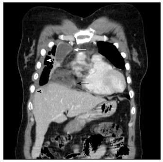
Figure 1 Chest computed tomography of the patient.
The black arrow indicates mediastinitis with fat lysis due to pseudocyst rupture. The white arrow indicates loculated effusion in the right anterior paramediastinal space. The black arrowhead indicates pleural effusion and the white arrowhead indicates pneumonic infiltration of the paracardiac area.
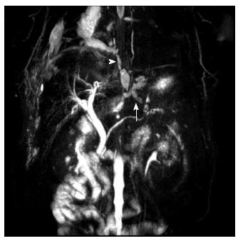
Figure 2 Magnetic resonance cholangiopancreatography revealed an 8-mm pseudocyst at the pancreatic tail connected with the distal pancreatic duct (arrow).
This pseudocyst communicated with another 2.6-cm pseudocyst in the right paraesophageal area. This second pseudocyst ruptured at the paraesophageal hiatus and extended to the right paracardiac area in the right middle mediastinum (arrowhead).
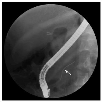
Figure 3 Endoscopic retrograde cholangiopancreatography of the patient.
The white arrow indicates that the guide wire did not pass through the mid-pancreatic area.
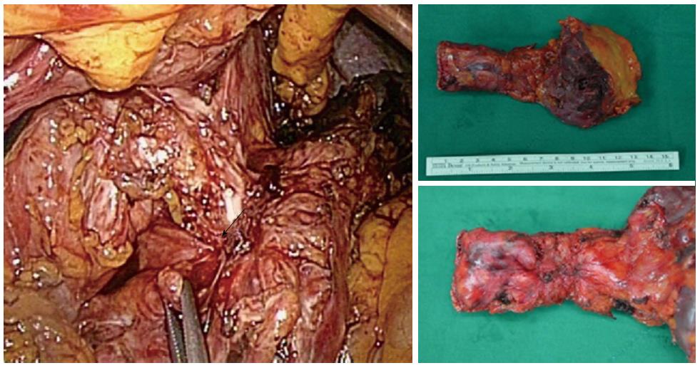
Figure 4 Laparoscopic distal pancreatectomy revealed severe adhesion due to chronic pancreatitis at the distal pancreas and stomach.
Fistula formation and pseudocyst rupture were seen. Adhesiolysis and fistula resection were performed.
- Citation: Choe IS, Kim YS, Lee TH, Kim SM, Song KH, Koo HS, Park JH, Pyo JS, Kim JY, Choi IS. Acute mediastinitis arising from pancreatic mediastinal fistula in recurrent pancreatitis. World J Gastroenterol 2014; 20(40): 14997-15000
- URL: https://www.wjgnet.com/1007-9327/full/v20/i40/14997.htm
- DOI: https://dx.doi.org/10.3748/wjg.v20.i40.14997


