Copyright
©2014 Baishideng Publishing Group Inc.
World J Gastroenterol. Oct 28, 2014; 20(40): 14855-14864
Published online Oct 28, 2014. doi: 10.3748/wjg.v20.i40.14855
Published online Oct 28, 2014. doi: 10.3748/wjg.v20.i40.14855
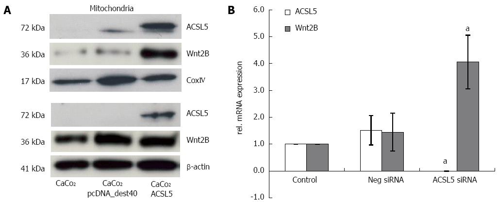
Figure 1 Expression of acyl-coA synthetase 5 and Wnt2B in mitochondrion, cytoplasm, and nucleus.
A: Western blot of mitochondrial and cytoplasmatic CaCo2 cell fractions, fraction specific reference antibodies were β-actin (cytoplasm) and CoxIV (mitochondria); B: Wnt2B mRNA expression in the presence or absence of acyl-CoA synthetase 5 (ACSL5) was analyzed by realtime PCR and normalized for the reference gene cyclophilin. aP < 0.05 vs control.
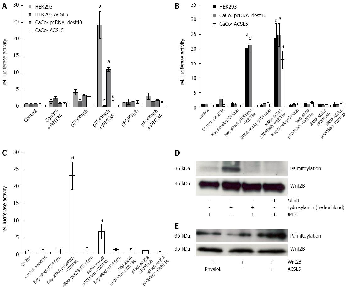
Figure 2 Wnt activation dependent on acyl-CoA synthetase 5 expression.
A: Luciferase reporter assay in HEK293 and CaCo2 cells stably or transiently transfected with acyl-CoA synthetase 5 (ACSL5) and transiently transfected with pTOPflash or pFOPflash. After activation with Wnt3A, Tcf reporter-derived luciferase activity was detected luminometric; B: Luciferase reporter assay after knockdown of ACSL5 by siRNA in HEK293 and CaCo2 cells; C: Luciferase reporter assay after knockdown of Wnt2B by siRNA in HEK293 cells; D: Palmitoylation of Wnt2B dependent on ACSL5 expression. Isolated mitochondria from HEK293 cells, transiently transfected and immunoprecipitated with Wnt2B, with or without treatment with palmostatin B, Hydroxylamine (hydrochloride), and BMCC. As a reference, Wnt2B protein expression was determined; E: Isolated mitochondria from CaCo2 cells, transiently transfected with Wnt2B and ACSL5, immunoprecipitated with Wnt2B, after treatment with palmostatin B, hydroxylamine (hydrochloride), and BMCC. aP < 0.05 vs control.
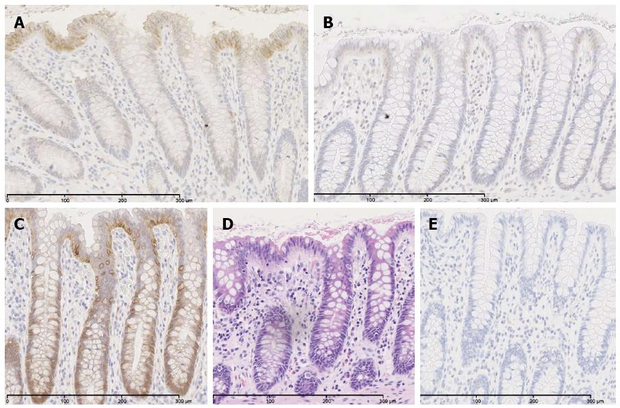
Figure 3 Expression of acyl-CoA synthetase 5, Wnt2B, and β-catenin in normal human intestinal mucosa.
Immunohistological staining of acyl-CoA synthetase 5 (A), Wnt2B (B), and β-catenin (C) on paraffin sections of normal human intestinal mucosa. For general view staining with hematoxylin and eosin (D), staining control (E).
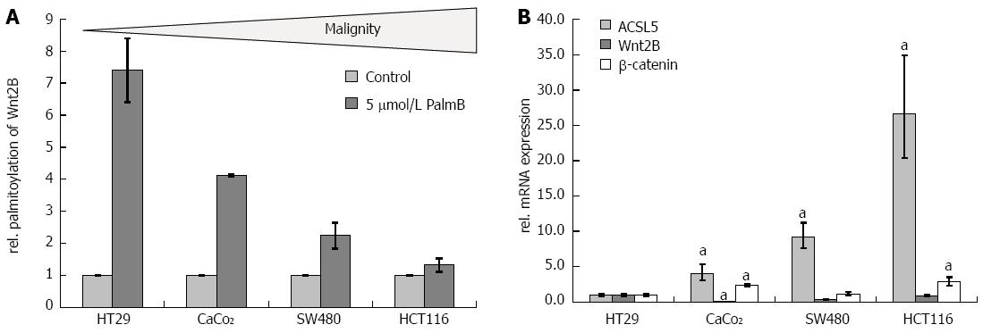
Figure 4 Palmitoylation of Wnt2B and expression levels of acyl-CoA synthetase 5, Wnt2B, and β-catenin in colon carcinoma cell lines of different malignancy statuses.
A: HT29, CaCo2, SW480, and HCT116 were treated with palmostatin B, mitochondria isolated, Wnt2B immunoprecipitated and palmitoylation was detected via streptavidin antibody in Western blot; B: mRNA expression of acyl-CoA synthetase 5 (ACSL5), Wnt2B, and β-catenin. Realtime PCR with cyclophilin as reference gene. aP < 0.05 vs control.
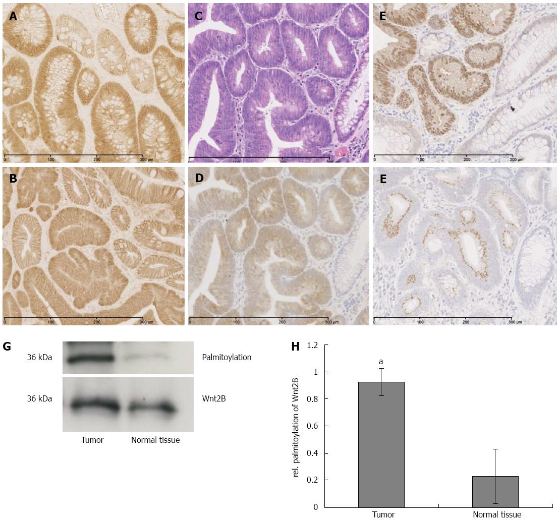
Figure 5 Proliferation and Wnt activation in adenoma of APCmin/+ mice and human familial adenomatous polyposis.
A: β-catenin staining on adenoma of human familial adenomatous polyposis and in APCmin/+ mice; B: Expression of acyl-CoA synthetase 5 (ACSL5), Wnt2B and β-catenin and palmitoylation of Wnt2B in human sporadic adenocarcinoma; C: General view staining with HE; D-F: Immunohistochemical staining with ACSL5, β-catenin, Wnt2B; G: Palmitoylation of Wnt2B in isolated mitochondria of human tissue, immunoprecipitated with Wnt2B, after treatment with palmostatin B, hydroxylamine (hydrochloride), and BMCC. Wnt2B as reference; H: Densitometry calculation of (G) with ImageJ. aP < 0.05 vs control.
- Citation: Klaus C, Schneider U, Hedberg C, Schütz AK, Bernhagen J, Waldmann H, Gassler N, Kaemmerer E. Modulating effects of acyl-CoA synthetase 5-derived mitochondrial Wnt2B palmitoylation on intestinal Wnt activity. World J Gastroenterol 2014; 20(40): 14855-14864
- URL: https://www.wjgnet.com/1007-9327/full/v20/i40/14855.htm
- DOI: https://dx.doi.org/10.3748/wjg.v20.i40.14855


