Copyright
©2014 Baishideng Publishing Group Co.
World J Gastroenterol. Jan 28, 2014; 20(4): 1123-1126
Published online Jan 28, 2014. doi: 10.3748/wjg.v20.i4.1123
Published online Jan 28, 2014. doi: 10.3748/wjg.v20.i4.1123
Figure 1 Images for preoperative diagnosis.
A: Upper gastrointestinal series demonstrated luminal narrowing and displacement of the duodenum by the mass (arrow head); B, C: Coronal reconstruction computed tomography image and coronal thick-slab T2-weighted image showed a multiloculated cystic mass (arrows) between the head of the pancreas and duodenum. The mass seemed to originate from the uncinate process of the pancreas, and the second and third portions of the duodenum were inferolaterally displaced and compressed by the mass (arrow heads).
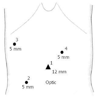
Figure 2 Laparoscopic port insertion site.
1: 12-mm optical umbilical port; 2, 5-mm main working port; 3: 5-mm port for liver retraction.
Figure 3 Laparoscopic procedures and intraoperative findings.
A: Laparoscopic intraoperative findings of duodenal duplication cyst after Kocher’s maneuver. Duplication cyst (white arrow heads) and mesenteric side of posterior wall of duodenum (black arrow heads); B: Demarcation of the mass surface after adhesiolysis. An arterial branch from the gastroduodenal artery supporting the mass was also noticed; C: Resection line with harmonic scalpel (white line).
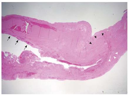
Figure 4 Pathological findings of resected specimen showing duodenal wall with partially denuded epithelium (hematoxylin and eosin staining, ×12.
5). The mucosal lining (black arrows), smooth muscle coat (black arrowheads), and glands (white arrowheads) were noticed.
- Citation: Byun J, Oh HM, Kim SH, Kim HY, Jung SE, Park KW, Kim WS. Laparoscopic partial cystectomy with mucosal stripping of extraluminal duodenal duplication cysts. World J Gastroenterol 2014; 20(4): 1123-1126
- URL: https://www.wjgnet.com/1007-9327/full/v20/i4/1123.htm
- DOI: https://dx.doi.org/10.3748/wjg.v20.i4.1123


