Copyright
©2014 Baishideng Publishing Group Co.
World J Gastroenterol. Jan 28, 2014; 20(4): 1005-1020
Published online Jan 28, 2014. doi: 10.3748/wjg.v20.i4.1005
Published online Jan 28, 2014. doi: 10.3748/wjg.v20.i4.1005
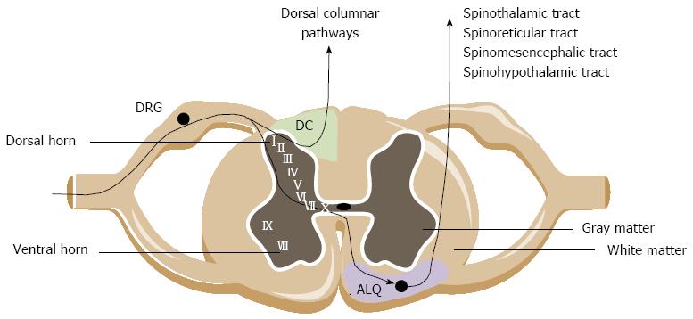
Figure 1 Cross section of the spinal cord.
The central branches of the visceral afferents innervating the lower gastrointestinal tract travel via the dorsal root ganglia (DRG) and project onto the second order neurons in laminae I, II, V, X of the spinal gray matter. Ascending pathways arise in the anterolateral quadrant (ALQ; purple zone) and the dorsal column (DC; green zone) region in the spinal cord and project to higher brain centers (, medulla, thalamus).
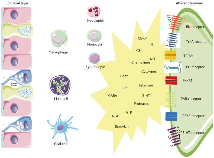
Figure 2 Scheme is oversimplified and limited to the cell types and mediators discussed in this review and represents a subset of cells and inflammatory mediators responsible for activation of gut sensory afferents after an initial inflammatory response.
5-HT: 5-hydroxytryptamine; BK: Bradykinin; CGRP: Calcitonin-gene-related peptide; ECC: Enterochromaffin cell; GABA: Gamma-amino butyric acid; NGF: Nerve growth factor; NO: Nitric oxide; PAR: Proteinase-activated receptor; PG: Prostaglandin; SP: Substance P; TrKA: Tyrosine receptor kinase A; TRPA1: Transient receptor potential ankyrin-1; TRPV1: Transient receptor potential vanilloid-1; P2X3: Purinergic P2X3 receptor.
- Citation: Vermeulen W, Man JGD, Pelckmans PA, Winter BYD. Neuroanatomy of lower gastrointestinal pain disorders. World J Gastroenterol 2014; 20(4): 1005-1020
- URL: https://www.wjgnet.com/1007-9327/full/v20/i4/1005.htm
- DOI: https://dx.doi.org/10.3748/wjg.v20.i4.1005


