Copyright
©2014 Baishideng Publishing Group Inc.
World J Gastroenterol. Oct 14, 2014; 20(38): 14058-14062
Published online Oct 14, 2014. doi: 10.3748/wjg.v20.i38.14058
Published online Oct 14, 2014. doi: 10.3748/wjg.v20.i38.14058
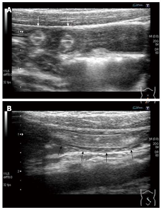
Figure 1 Abdominal ultrasound examination at the pediatric emergency room.
A: Ultrasonography shows multiple, round-shaped structures with a target-like appearance within the small intestine (white arrows); B: In a longitudinal scan of the small intestine, ultrasound shows a tubular structure with two parallel, outer high echogenic lines (black arrows). Space between the intestinal wall and the tubular structure was not observed.
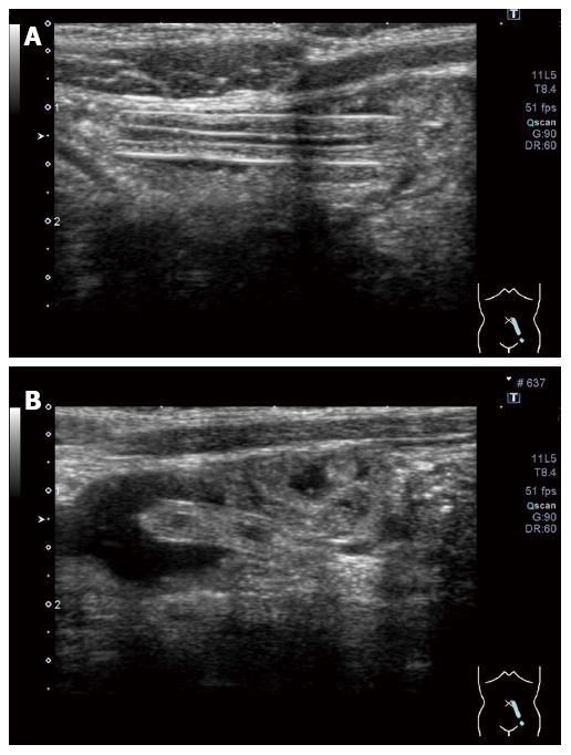
Figure 2 Abdominal ultrasound examination on the day after admission.
The tubular structure in the patient’s small intestine was still observed. An ultrasound examination shows a space between the intestinal wall and the tubular structure, and fluid could pass through this space.
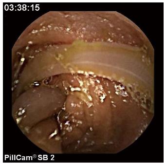
Figure 3 Capsule endoscopy.
Capsule endoscopy shows a single roundworm moving in the jejunum. There appears to be more than one worm body. However, movement of each part of a body able to be visualized was coordinated. A single worm of Ascaris infection was diagnosed.
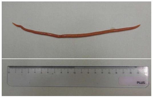
Figure 4 Excreted Ascaris lumbricoides after treatment.
A single worm of Ascaris lumbricoides was excreted the day after treatment of pyrantel pamoate. The worm length was approximately 20 cm.
- Citation: Umetsu S, Sogo T, Iwasawa K, Kondo T, Tsunoda T, Oikawa-Kawamoto M, Komatsu H, Inui A, Fujisawa T. Intestinal ascariasis at pediatric emergency room in a developed country. World J Gastroenterol 2014; 20(38): 14058-14062
- URL: https://www.wjgnet.com/1007-9327/full/v20/i38/14058.htm
- DOI: https://dx.doi.org/10.3748/wjg.v20.i38.14058


