Copyright
©2014 Baishideng Publishing Group Inc.
World J Gastroenterol. Oct 14, 2014; 20(38): 14004-14009
Published online Oct 14, 2014. doi: 10.3748/wjg.v20.i38.14004
Published online Oct 14, 2014. doi: 10.3748/wjg.v20.i38.14004
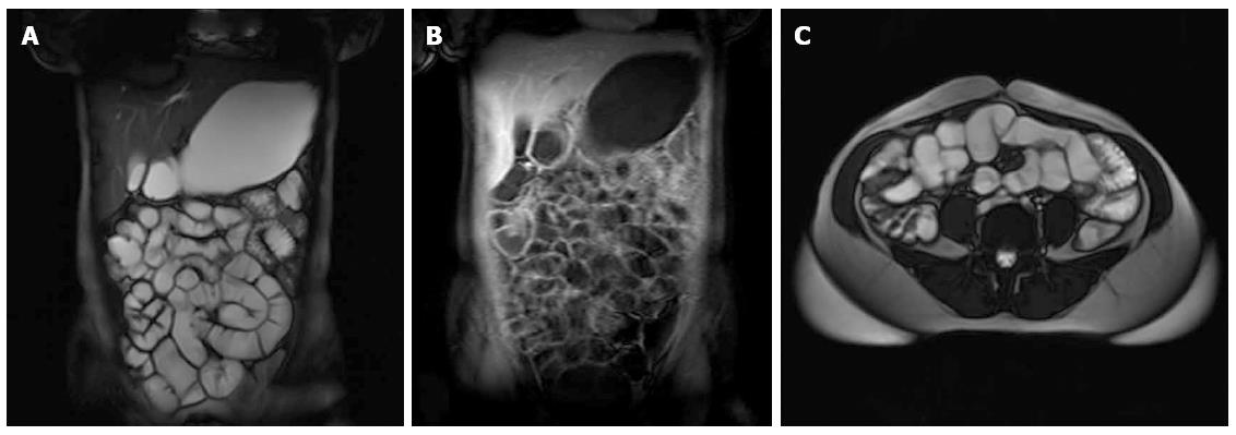
Figure 1 Normal magnetic resonance enterography findings.
A: Coronal T2 fast imaging with steady-state precession (FISP); B: Coronal T1 volumetric interpolated breath-hold examination with contrast; C: Axial T2 FISP images.
Figure 2 Magnetic resonance enterography findings of antral gastritis.
A: Axial T2 half-Fourier acquisition single-shot turbo spin-echo; B: Axial T1 volumetric interpolated breath-hold examination prior to contrast; C: Axial T1 VIBE after contrast injection. Antrum is thickened (arrows) while periantral soft tissues remain unaffected.
Figure 3 Magnetic resonance enterography findings of adenocarcinoma of the duodenum.
A: Axial T2 half-fourier acquisition single-shot turbo spin-echo; B: Axial T1 volumetric interpolated breath-hold examination (VIBE) with contrast; C: Coronal T2 fast imaging with steady-state precession; D: Coronal T1 VIBE with contrast. Asymmetric thickening of the duodenal wall and contrast enhancement is seen with luminal narrowing in the proximal and distal ends with slight dilatation in the center (arrows). Periduodenal mesenteric adipose tissue appears infiltrated, and the tumor is in close relation with the abdominal aorta (arrowheads).
Figure 4 Magnetic resonance enterography findings suggesting Crohn’s disease.
A: Axial T2 half-Fourier acquisition single-shot turbo spin-echo (HASTE); B: Axial T1 volumetric interpolated breath-hold examination (VIBE) with contrast; C: Coronal T2 HASTE; D: Coronal T1 VIBE with contrast. Wall thickening and contrast enhancement in distal ileal segments (arrows) are observed. Ileoileal and ileomesenteric fistula formations (arrowheads) support the diagnosis of Crohn’s disease.
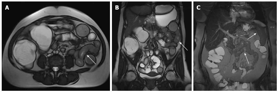
Figure 5 Atypical magnetic resonance enterography findings in a case with small bowel lymphoma.
A: Axial T2 fast imaging with steady-state precession (FISP); B: Coronal T2 FISP; C: T2 maximum intensity projection images. Long segment wall thickening and luminal narrowing in the jejunum (arrows) without accompanying lymphadenopathy. The biopsy showed an isolated small bowel lymphoma, though imaging findings were not strongly suggestive.
- Citation: Cengic I, Tureli D, Aydin H, Bugdayci O, Imeryuz N, Tuney D. Magnetic resonance enterography in refractory iron deficiency anemia: A pictorial overview. World J Gastroenterol 2014; 20(38): 14004-14009
- URL: https://www.wjgnet.com/1007-9327/full/v20/i38/14004.htm
- DOI: https://dx.doi.org/10.3748/wjg.v20.i38.14004


