Copyright
©2014 Baishideng Publishing Group Inc.
World J Gastroenterol. Oct 14, 2014; 20(38): 13705-13717
Published online Oct 14, 2014. doi: 10.3748/wjg.v20.i38.13705
Published online Oct 14, 2014. doi: 10.3748/wjg.v20.i38.13705
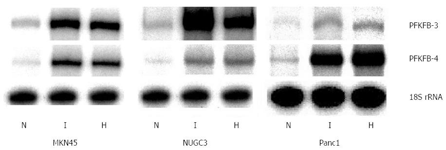
Figure 1 Effect of hypoxia (H) and hypoxia mimic dimethyloxalylglycine (I) on the expression of 6-phosphofructo-2-kinase/fructose-2,6-bisphosphatase-3 and -4 mRNA in human gastric cancer cell lines MKN45 and NUGC3 and pancreatic cancer cell line Panc1.
Measured by ribonuclease protection assay, N: Control (normoxic) cells[32]. PFKFB: 6-phosphofructo-2-kinase/fructose-2,6-bisphosphatase.
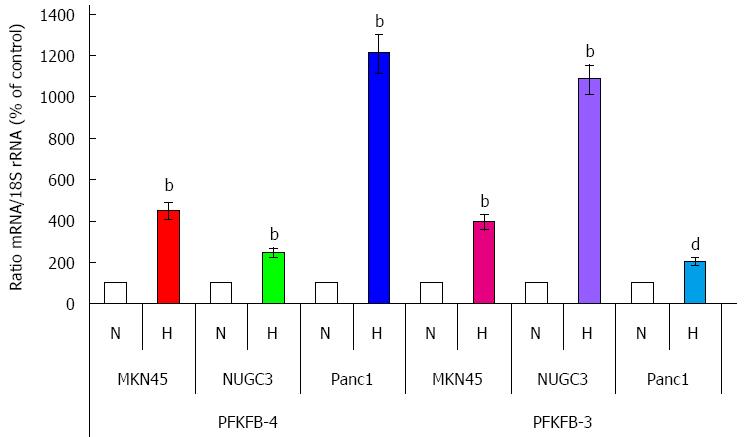
Figure 2 Quantification of ribonuclease protection assay of the effect of hypoxia (H) on the expression level of 6-phosphofructo-2-kinase/fructose-2,6-bisphosphatase-4 and -3 mRNAs in human gastric (MKN45 and NUGC3) and pancreatic (Panc1) cancer cell lines.
bP < 0.01 vs control cells; dP < 0.01 vs control cells[32]. N: Normoxic (control) cells. PFKFB: 6-phosphofructo-2-kinase/fructose-2,6-bisphosphatase.
Figure 3 Western blot analysis of 6-phosphofructo-2-kinase/fructose-2,6-bisphosphatase-4 protein in human gastric (MKN45 and NUGC3) and pancreatic (Panc1) cancer cell lines: Effect of hypoxia (H) and dimethyloxalylglycine (I)[32].
PFKFB: 6-phosphofructo-2-kinase/fructose-2,6-bisphosphatase.
Figure 4 Expression of hypoxia inducible factor-1α protein (Western blotting; A) and hypoxia inducible factor-1α and hypoxia inducible factor-2α mRNA (ribonuclease protection assay; B) in human gastric (MKN45 and NUGC3) and pancreatic (Panc1) cancer cell lines: effect of hypoxia (H) and dimethyloxalylglycine (I)[32].
HIF: Hypoxia inducible factor.
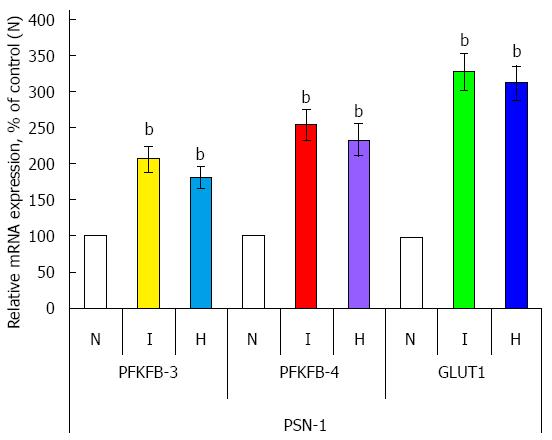
Figure 5 Effect of hypoxia (H) and hypoxia mimic dimethyloxalylglycine (I) on the expression level of 6-phosphofructo-2-kinase/fructose-2,6-bisphosphatase-3, -4, and GLUT1 mRNAs (measured by qPCR) in human pancreatic (PSN-1) cancer cells.
n = 4; bP < 0.01 vs control cells. N: Normoxic (control) cells; PFKFB: 6-phosphofructo-2-kinase/fructose-2,6-bisphosphatase.
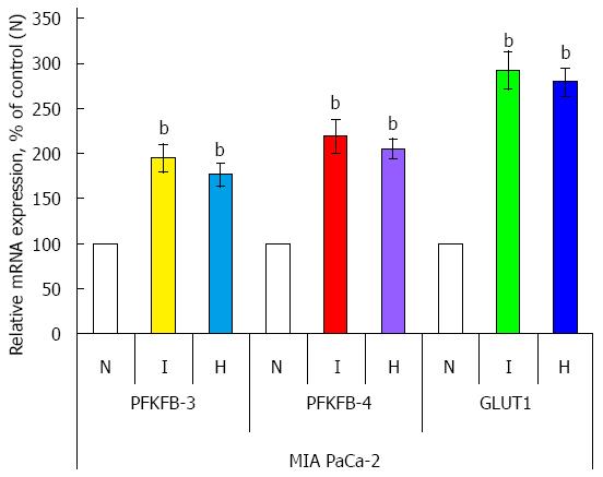
Figure 6 Effect of hypoxia (H) and hypoxia mimic dimethyloxalylglycine (I) on the expression level of 6-phosphofructo-2-kinase/fructose-2,6-bisphosphatase-3, -4, and GLUT1 mRNAs (measured by qPCR) in human pancreatic (MIA PaCa-2) cancer cells.
n = 4, bP < 0.01 vs control cells. N: Normoxic (control) cells; PFKFB: 6-phosphofructo-2-kinase/fructose-2,6-bisphosphatase.
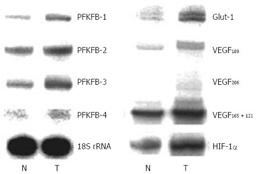
Figure 7 Representative polyacrylamide gel autoradiograph employed in a typical ribonuclease protection assay of different 6-phosphofructo-2-kinase/fructose-2,6-bisphosphatase genes (PFKFB-1, PFKFB-2, PFKFB-3, and PFKFB-4), GLUT1, hypoxia inducible factor-1α, and different alternative splice variants of VEGF-A in gastric malignant tumors (T) and non-malignant tissue counterparts (N) from same patients.
The 18S rRNA expressions were used as control of RNA quantity used for analysis[32]. HIF: Hypoxia inducible factor; PFKFB: 6-phosphofructo-2-kinase/fructose-2,6-bisphosphatase.
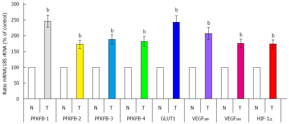
Figure 8 Quantification of ribonuclease protection assay of 6-phosphofructo-2-kinase/fructose-2,6-bisphosphatase-1, 6-phosphofructo-2-kinase/fructose-2,6-bisphosphatase-2, 6-phosphofructo-2-kinase/fructose-2,6-bisphosphatase-3, 6-phosphofructo-2-kinase/fructose-2,6-bisphosphatase-4, GLUT1, hypoxia inducible factor-1α, and splice variants of VEGF-A mRNA in human gastric malignant tumors (T) and corresponding non-malignant tissue (N) from the same patients.
bP < 0.01 vs control cells[32]. HIF: Hypoxia inducible factor; PFKFB: 6-phosphofructo-2-kinase/fructose-2,6-bisphosphatase; VEGF: Vascular endothelial growth factor.
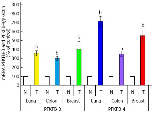
Figure 9 Quantification of ribonuclease protection assay of 6-phosphofructo-2-kinase/fructose-2,6-bisphosphatase-3 and 6-phosphofructo-2-kinase/fructose-2,6-bisphosphatase-4 mRNA expressions in lung, colon, and breast malignant tumors (T) and corresponding non-malignant tissue counterparts (C).
Values of PFKFB-3 and PFKFB-4 mRNA expressions were normalized to 18S rRNA; n = 15-20, bP < 0.01 vs non-malignant tissues[30,33]. PFKFB: 6-phosphofructo-2-kinase/fructose-2,6-bisphosphatase.
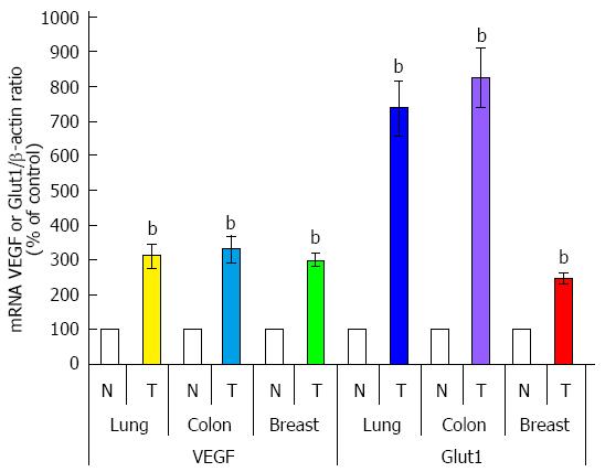
Figure 10 Quantification of ribonuclease protection assay of VEGF and Glut1 mRNA expressions in lung, colon, and breast malignant tumors (T) and corresponding non-malignant tissue counterparts (C).
Values of VEGF and Glut1 mRNA expressions were normalized to 18S rRNA; n = 15-20; bP < 0.01 vs non-malignant tissues[30,33]. PFKFB: 6-phosphofructo-2-kinase/fructose-2,6-bisphosphatase; VEGF: Vascular endothelial growth factor.
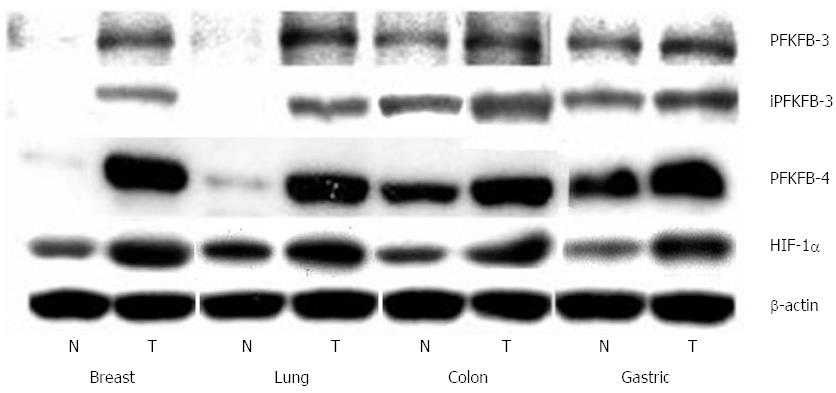
Figure 11 Representative Western blot analysis of 6-phosphofructo-2-kinase/fructose-2,6-bisphosphatase-3, inducible 6-phosphofructo-2-kinase/fructose-2,6-bisphosphatase-3, 6-phosphofructo-2-kinase/fructose-2,6-bisphosphatase-4, and hypoxia inducible factor-1α protein levels in breast, lung, colon, and stomach malignant tumors (T) and non-malignant (control) tissues counterparts (N) from same patients.
The actin was used to ensure equal loading of the sample[30,32,33]. HIF: Hypoxia inducible factor; PFKFB: 6-phosphofructo-2-kinase/fructose-2,6-bisphosphatase.
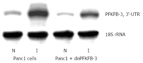
Figure 12 Representative polyacrylamide gel autoradiograph employed in a typical ribonuclease protection assay of endogenous 6-phosphofructo-2-kinase/fructose-2,6-bisphosphatase-3 mRNA in pancreatic carcinoma cell line Panc1, stable transfected by pcDNA3.
1(+) vector (Panc1 cells) or by dominant/negative 6-phosphofructo-2-kinase/fructose-2,6-bisphosphatase-3 (Panc1 + dnPFKFB-3) in normoxic (N) condition and after treatment of Panc1 cells with dimethyloxalylglycine, inhibitor of prolyl hydroxylase (I; 1 mmol/L for 6 h). The 18S rRNA antisense probe was used as control of analyzed RNA quantity[89]. PFKFB: 6-phosphofructo-2-kinase/fructose-2,6-bisphosphatase.
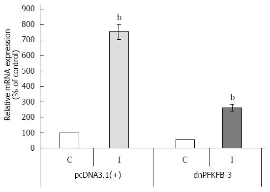
Figure 13 Quantification of ribonuclease protection assay of endogenous 6-phosphofructo-2-kinase/fructose-2,6-bisphosphatase-3 mRNA expression in pancreatic carcinoma cell line Panc1, stable transfected by pcDNA3.
1(+) vector or dominant/negative 6-phosphofructo-2-kinase/fructose-2,6-bisphosphatase-3 in normoxic (control) condition (C) and after treatment of Panc1 cells with dimethyloxalylglycine, inhibitor of prolyl hydroxylase (I). n = 5; bP < 0.01 vs control[89]. dnPFKFB-3: Dominant/negative 6-phosphofructo-2-kinase/fructose-2,6-bisphosphatase-3.
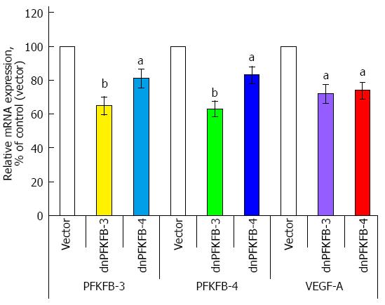
Figure 14 Endogenous 6-phosphofructo-2-kinase/fructose-2,6-bisphosphatase-3, 6-phosphofructo-2-kinase/fructose-2,6-bisphosphatase-4, and vascular endothelial growth factor mRNA expressions in pancreatic carcinoma cell line PSN-1, stable transfected with pcDNA3.
1(+) vector (Vector), dominant/negative 6-phosphofructo-2-kinase/fructose-2,6-bisphosphatase-3, and dominant/negative 6-phosphofructo-2-kinase/fructose-2,6-bisphosphatase-4, measured by ribonuclease protection assay. n = 5; aP < 0.05 vs control; bP < 0.01 vs control[89]. PFKFB: 6-phosphofructo-2-kinase/fructose-2,6-bisphosphatase; VEGF: Vascular endothelial growth factor.
- Citation: Minchenko OH, Tsuchihara K, Minchenko DO, Bikfalvi A, Esumi H. Mechanisms of regulation of PFKFB expression in pancreatic and gastric cancer cells. World J Gastroenterol 2014; 20(38): 13705-13717
- URL: https://www.wjgnet.com/1007-9327/full/v20/i38/13705.htm
- DOI: https://dx.doi.org/10.3748/wjg.v20.i38.13705


