Copyright
©2014 Baishideng Publishing Group Inc.
World J Gastroenterol. Oct 7, 2014; 20(37): 13615-13619
Published online Oct 7, 2014. doi: 10.3748/wjg.v20.i37.13615
Published online Oct 7, 2014. doi: 10.3748/wjg.v20.i37.13615
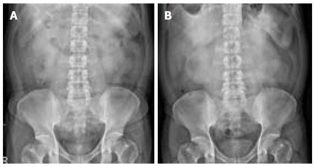
Figure 1 Serial abdominal radiographs showing gas within the small bowel overlying the liver and relative radiolucencies lateral to the liver, suggesting intestinal incarceration.
The intestinal diverticulum between the diaphragm and liver is prominent in the area of the latter. A: 5 mo ago; B: During the acute episode.
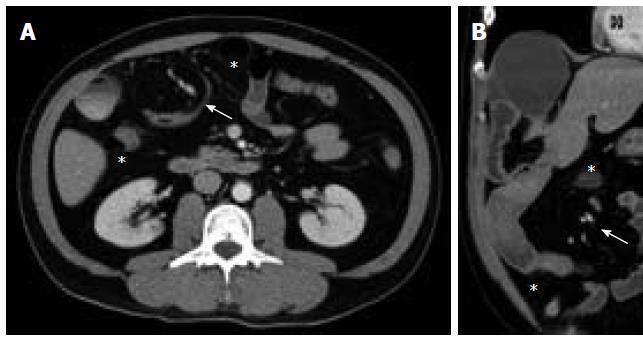
Figure 2 Computed tomography scans showing the incarceration of Meckel’s diverticulum and the small bowel in the right subphrenic space, causing the depression of liver surface.
The twisted mesenteric vessels (arrow) are indicative of a volvulus; the group of small-bowel loops lateral to the colon (asterisk) also suggest a transmesocolic internal hernia. A: Axial view; B: Coronal view.
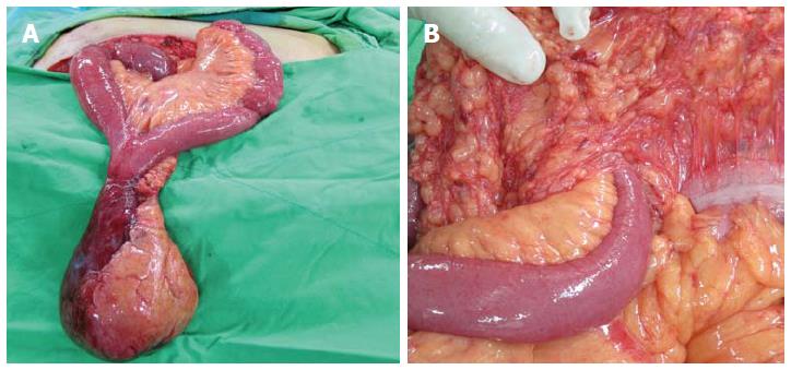
Figure 3 Intraoperative photographs.
A: The herniated Meckel’s diverticulum and small bowel, showing the completion of the manual reduction of the mesenteric volvulus; B: The inlet of the internal hernia in the transverse mesocolon.
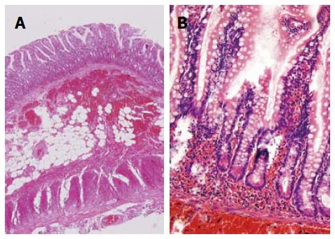
Figure 4 Hematoxylin eosin stains of the resected diverticulum showing mucosal infarction, extensive submucosal hemorrhage, and inflammatory cell infiltration of the tissues of the ileum and continuous muscle layer, suggesting acute bowel ischemia.
A: × 25; B: ×200.
- Citation: Wu SY, Ho MH, Hsu SD. Meckel's diverticulum incarcerated in a transmesocolic internal hernia. World J Gastroenterol 2014; 20(37): 13615-13619
- URL: https://www.wjgnet.com/1007-9327/full/v20/i37/13615.htm
- DOI: https://dx.doi.org/10.3748/wjg.v20.i37.13615


