Copyright
©2014 Baishideng Publishing Group Inc.
World J Gastroenterol. Sep 21, 2014; 20(35): 12682-12686
Published online Sep 21, 2014. doi: 10.3748/wjg.v20.i35.12682
Published online Sep 21, 2014. doi: 10.3748/wjg.v20.i35.12682
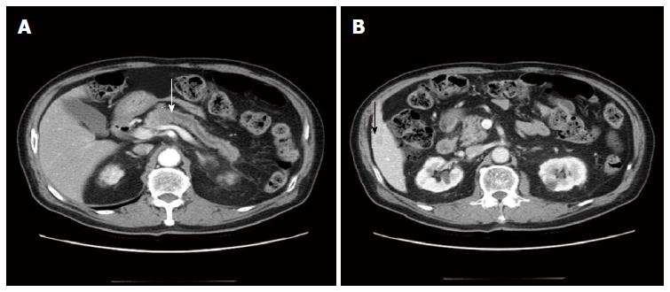
Figure 1 Axial pancreas-enhanced computed tomography findings.
A: A 2.2 cm low attenuated mass in the pancreas body with diffuse pancreas duct dilatation (white arrow: pancreatic head mass); B: A 1.3 cm low attenuated hepatic nodule in S5 (black arrow: hepatic mass).
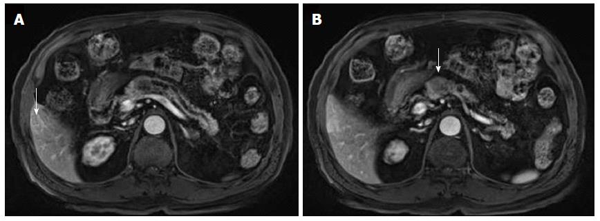
Figure 2 Magnetic resonance imaging findings.
A: A T1 weighted magnetic resonance imaging (MRI) revealed a 2.2 cm mild low signal intensity pancreatic body mass with diffuse distal pancreatic duct dilatation (arrow: hepatic mass in S5); B: An MRI in the arterial phase revealed a 1.3 cm round, peripheral enhanced mass in S5 of the liver (arrow: pancreatic head mass).
Figure 3 Pancreatic neoplasia biopsy.
A: Dual disparate sarcomatous (right) and carcinomatous (left) components of the tumor [hematoxylin and eosin (HE) stain, × 100); B: Sarcomatous component (HE stain, × 400); C: Carcinomatous component (HE stain, × 200).
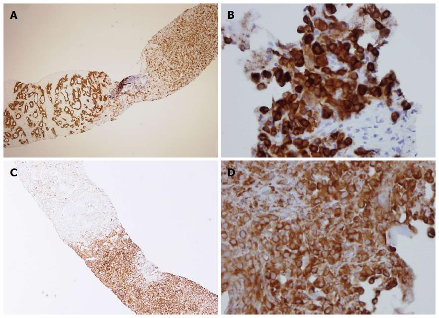
Figure 4 Immunohistopathology of the pancreatic tumor.
A: A pancreatic biopsy revealed a sarcomatoid component (right) containing mesenchymal tumor cells and a carcinomatous component that gave rise to tumor glands (left) [pan-cytokeratin (CK) staining, × 40]; B: The sarcomatoid component from the liver was also positive for pan-CK (× 400); C: In the pancreatic biopsy, the sarcomatous component stained positive for vimentin (right) while the adenomatous component was negative for vimentin (left) (× 40); D: The sarcomatoid component from the liver stained positive for vimentin (× 400).
- Citation: Kim HS, Kim JI, Jeong M, Seo JH, Kim IK, Cheung DY, Kim TJ, Kang CS. Pancreatic adenocarcinosarcoma of monoclonal origin: A case report. World J Gastroenterol 2014; 20(35): 12682-12686
- URL: https://www.wjgnet.com/1007-9327/full/v20/i35/12682.htm
- DOI: https://dx.doi.org/10.3748/wjg.v20.i35.12682


