Copyright
©2014 Baishideng Publishing Group Inc.
World J Gastroenterol. Sep 14, 2014; 20(34): 12341-12345
Published online Sep 14, 2014. doi: 10.3748/wjg.v20.i34.12341
Published online Sep 14, 2014. doi: 10.3748/wjg.v20.i34.12341
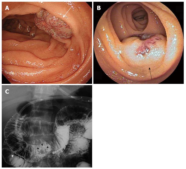
Figure 1 Hemangiomas in the duodenum and jejunum.
A: Upper gastrointestinal endoscopy shows a hemangioma of the duodenum in the third portion (white arrow); B: Double balloon endoscopy shows a hemangioma of the jejunum (black arrow); C: A barium study shows the duodenal hemangioma (black arrowheads) located in the third portion of duodenum, approximately 4.0 cm from the anal side of the inferior flexure (white arrowheads).
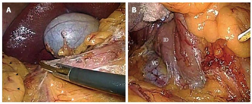
Figure 2 Laparoscopic Kocher maneuver.
A: Right colon (black arrowheads) and mesocolon (white arrowheads) were freed laparoscopically from the superior flexure of the duodenum (SD). B: Dissection around the inferior flexure of the duodenum (ID) was extended beyond the vena cava (VC).
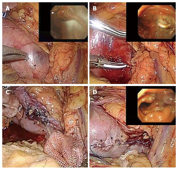
Figure 3 Laparoscopic resection of the duodenal hemangioma.
A: Intraoperative gastroduodenal endoscopy shows the intraluminal location of the hemangioma (white arrowheads), corresponding with the location identified by laparoscopy with the serosal color change of the duodenum (black arrowheads); B: Resection of the lesion was performed using ultrasonic coagulating shears with a laparo-endoscopic view; C: The defect of the duodenal wall was closed manually using an intracorporeal running suture in the whole layer. The suture line (black arrowheads) was extremely close to the pancreas (white arrowheads); D: After the intracorporeal running suture was placed in the seromusclar layer, the duodenum was reinflated during the laparoscopy to detect any bleeding or leakage from the suturing line (black arrowheads) and to confirm a patent lumen by intraluminal endoscopy.
- Citation: Kanaji S, Nakamura T, Nishi M, Yamamoto M, Kanemitu K, Yamashiita K, Imanishi T, Sumi Y, Suzuki S, Tanaka K, Kakeji Y. Laparoscopic partial resection for hemangioma in the third portion of the duodenum. World J Gastroenterol 2014; 20(34): 12341-12345
- URL: https://www.wjgnet.com/1007-9327/full/v20/i34/12341.htm
- DOI: https://dx.doi.org/10.3748/wjg.v20.i34.12341


