Copyright
©2014 Baishideng Publishing Group Inc.
World J Gastroenterol. Sep 7, 2014; 20(33): 11910-11915
Published online Sep 7, 2014. doi: 10.3748/wjg.v20.i33.11910
Published online Sep 7, 2014. doi: 10.3748/wjg.v20.i33.11910
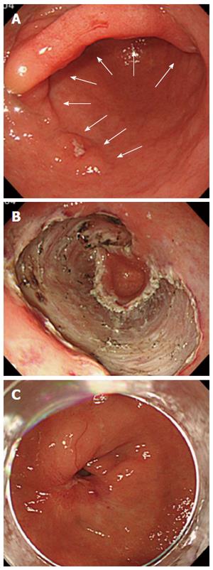
Figure 1 We treated a case of severe stenosis in the antrum after the endoscopic submucosal dissection procedure.
The circumferential extent of the mucosal defect was > 3/4. A: The white arrow shows that the lesion constituted of a semi-circumferential, laterally spreading type 0-IIc early gastric cancer on the anterior wall of the antrum; B: The circumferential extent of the mucosal defect was > 4/5 after the performance of the ESD procedure; C: Three months after the endoscopic submucosal dissection operation, the patient experienced a severe stricture of the gastric antrum. Subsequently, we had to perform a mucosal incision and local steroid injection.
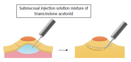
Figure 2 Schematic diagram of our proposed new method.
Schematic diagram of our proposed new method consists of submucosal injection of a mixture of solution containing triamcinolone acetonide 8 mL, glycerol 16 mL, and hyaluronic acid 16 mL, in addition to a small amount of indigo carmine and epinephrine, which was injected during the performance of the endoscopic submucosal dissection (ESD). The lesions were treated by standard ESD procedures.
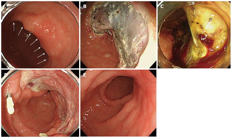
Figure 3 We performed our proposed new method during endoscopic submucosal dissection at gastric antrum (Case 1).
A: Endoscopic imaging showed that the lesion constituted a semi-circumferential, laterally spreading type 0-IIc early gastric cancer in the posterior wall of the antrum (white arrow); B: A three-fourths circumferential mucosa in the antrum was removed by standard endoscopic submucosal dissection (ESD) procedure; C: The day after the ESD operation, there was abundant vascularization in the ESD ulcer; D: Two weeks after the ESD operation, there was more granulation tissue than in a typical ESD ulcer; E: Three months after the ESD operation, there were no strictures concomitant with the healing process near the ESD ulcer.
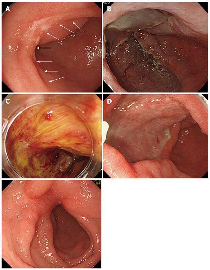
Figure 4 We performed our proposed new method during endoscopic submucosal dissection at gastric antrum (Case 2).
A: Endoscopic imaging showed that the lesion constituted a one-third circumferential, laterally spreading type 0-IIc early gastric cancer on the anterior wall of the antrum (white arrow); B: A three-fourths circumferential mucosa in the antrum was removed by standard endoscopic submucosal dissection (ESD) procedure; C: The day after the ESD, there was abundant vascularization in the ESD ulcer; D: Two weeks after the ESD operation, there was more granulation tissue than usual in the ESD ulcer; E: Three months after the ESD operation, there were no strictures concomitant with the healing process near the ESD ulcer.
- Citation: Nishiyama N, Mori H, Kobara H, Rafiq K, Fujihara S, Matsunaga T, Ayaki M, Yachida T, Oryu M, Masaki T. Novel method to prevent gastric antral strictures after endoscopic submucosal dissection: Using triamcinolone. World J Gastroenterol 2014; 20(33): 11910-11915
- URL: https://www.wjgnet.com/1007-9327/full/v20/i33/11910.htm
- DOI: https://dx.doi.org/10.3748/wjg.v20.i33.11910


