Copyright
©2014 Baishideng Publishing Group Inc.
World J Gastroenterol. Aug 14, 2014; 20(30): 10478-10485
Published online Aug 14, 2014. doi: 10.3748/wjg.v20.i30.10478
Published online Aug 14, 2014. doi: 10.3748/wjg.v20.i30.10478
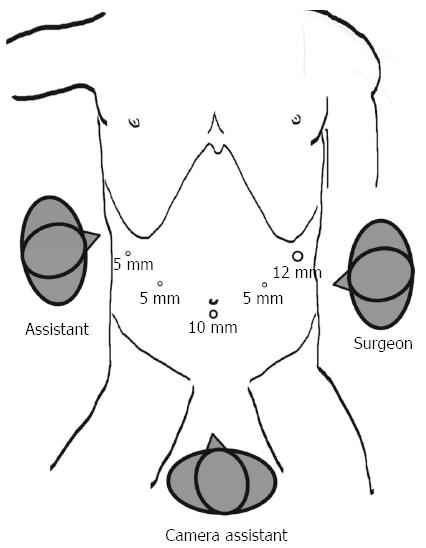
Figure 1 Trocar placements for totally laparoscopic distal gastrectomy.
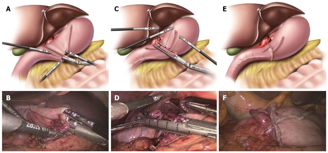
Figure 2 Conventional delta-shaped gastroduodenostomy procedure.
A: Diagram showing that the 60-mm endoscopic linear stapler was positioned to join the posterior walls together; B: Intraoperative image showing that the 60-mm endoscopic linear stapler was positioned to join the posterior walls together; C: Diagram showing that three sutures were added to each end of the common stab incision and cutting edges of the stomach and duodenum to obtain a better involution and pull; D: Intraoperative image showing that three sutures were added to each end of the common stab incision and cutting edges of the stomach and duodenum to obtain a better involution and pull; E: Diagram showing a completed conventional delta-shaped gastroduodenostomy; F: Intraoperative image showing a completed conventional delta-shaped gastroduodenostomy.
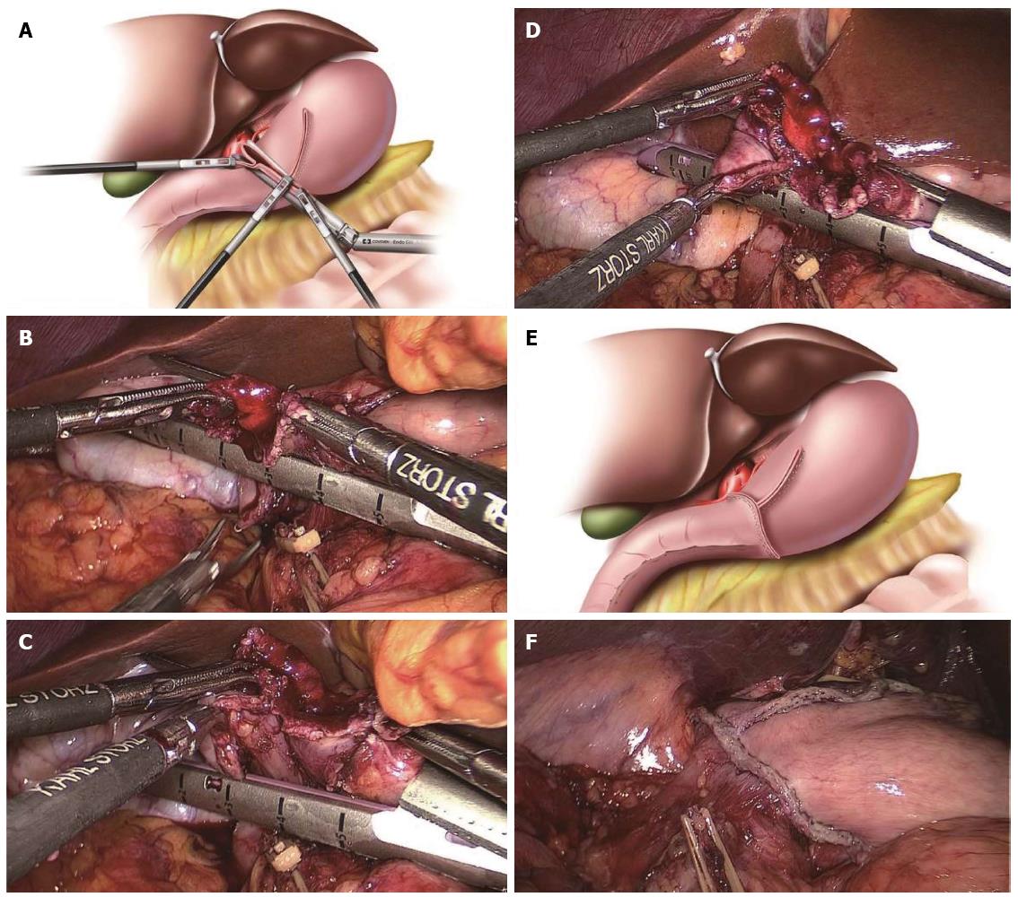
Figure 3 Modified delta-shaped gastroduodenostomy procedure.
A: Diagram showing the completed involution of the common stab incision using the instruments of the surgeon and assistant with the other end of the duodenal cutting edge being pulled into the stapler in the modified delta-shaped gastroduodenostomy; B: Intraoperative image showing the surgeon and assistant using their instruments to complete the initial involution of the common stab incision; C: Intraoperative image showing that the other end of the duodenal cutting edge was pulled up by the assistant’s right forceps; D: Intraoperative image showing that the other end of the duodenal cutting edge was pulled into the stapler; E: Diagram showing the completed inverted T-shaped appearance of the anastomosis; F: Intraoperative image showing the completed inverted T-shaped appearance of the anastomosis.
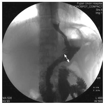
Figure 4 Upper gastrointestinal radiography film.
The upper gastrointestinal radiography film with diatrizoate meglumine as the contrast medium on postoperative day 7 for one patient who underwent a delta-shaped gastroduodenostomy. The inner diameter of the anastomosis was measured as the length of the white arrow as shown in the figure.
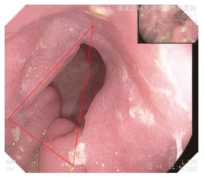
Figure 5 Gastroscopic image of one patient who underwent a delta-shaped gastroduodenostomy at 3 mo postoperatively.
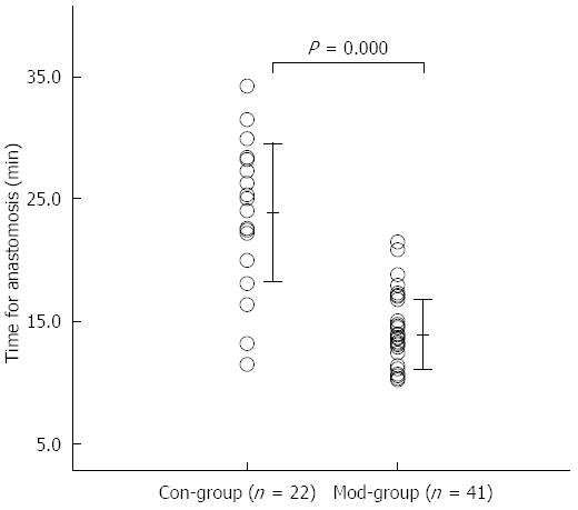
Figure 6 Effect of the modified delta-shaped gastroduodenostomy on the anastomosis time.
Comparison of the anastomosis time between the Con-Group and the Mod-Group. Con-Group: Conventional delta-shaped gastroduodenostomy group; Mod-Group: Modified version of the delta-shaped gastroduodenostomy group.
- Citation: Huang CM, Lin M, Lin JX, Zheng CH, Li P, Xie JW, Wang JB, Lu J. Comparision of modified and conventional delta-shaped gastroduodenostomy in totally laparoscopic surgery. World J Gastroenterol 2014; 20(30): 10478-10485
- URL: https://www.wjgnet.com/1007-9327/full/v20/i30/10478.htm
- DOI: https://dx.doi.org/10.3748/wjg.v20.i30.10478


