Copyright
©2014 Baishideng Publishing Group Co.
World J Gastroenterol. Jan 21, 2014; 20(3): 863-868
Published online Jan 21, 2014. doi: 10.3748/wjg.v20.i3.863
Published online Jan 21, 2014. doi: 10.3748/wjg.v20.i3.863
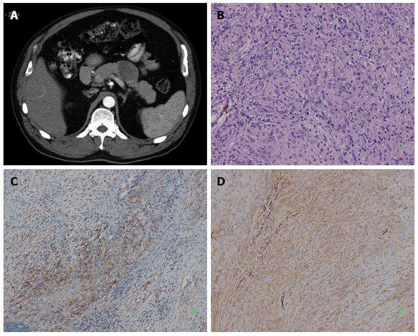
Figure 1 An abdominal mass was detected by ultrasound in a 61-year-old man with no clinical symptoms.
A: Enhanced abdominal computed tomography scan showed a solid mass of the pancreatic body, and the tumor located at pancreas tail next to splenic artery; B: The tumor was composed of spindle cell (HE, × 200); C: Immunoreactivity of the tumor cells for CD117 was positive (+) (SP × 100); D: Immunoreactivity of the tumor cells for CD34 was positive (++) (SP × 200).
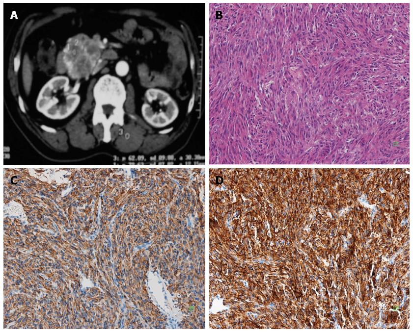
Figure 2 A 60-year-old man with no clinical symptoms underwent computed tomography.
A: Enhanced abdominal computed tomography scan showed a solid mass in the pancreatic head; B: The tumor was composed of spindle cell (HE, × 200); C: Immunoreactivity of the tumor cells for CD117 was positive (+) (SP × 200); D: Immunoreactivity of the tumor cells for DOG-1 was positive (+++) (SP × 200).
- Citation: Tian YT, Liu H, Shi SS, Xie YB, Xu Q, Zhang JW, Zhao DB, Wang CF, Chen YT. Malignant extra-gastrointestinal stromal tumor of the pancreas: Report of two cases and review of the literature. World J Gastroenterol 2014; 20(3): 863-868
- URL: https://www.wjgnet.com/1007-9327/full/v20/i3/863.htm
- DOI: https://dx.doi.org/10.3748/wjg.v20.i3.863


