Copyright
©2014 Baishideng Publishing Group Co.
World J Gastroenterol. Jan 21, 2014; 20(3): 852-856
Published online Jan 21, 2014. doi: 10.3748/wjg.v20.i3.852
Published online Jan 21, 2014. doi: 10.3748/wjg.v20.i3.852
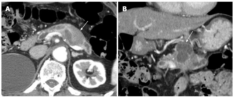
Figure 1 Abdominal computed tomography.
A: An axial section image of an abdominal computed tomography showed dilatation of the main pancreatic duct (arrow head) and the adjacent solid tumor (arrow); B: The coronal section image revealed a cystic lesion measuring 6.0 cm × 3.5 cm in the body and tail of the pancreas (arrow).
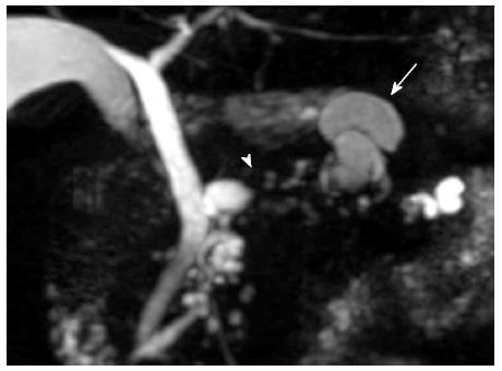
Figure 2 Magnetic resonance cholangiopancreatography revealed a segmental defect of the main pancreatic duct by the solid tumor (arrow head) associated with the multilocular cystic lesion (arrow) in the body and tail of the pancreas.
Figure 3 Endoscopic retrograde cholangiopancreatography showed the dilatation of the main pancreatic duct.
A: A filling defect in the main pancreatic duct (MPD) (arrow head); B: Intraductal ultrasonography demonstrated a filling lesion in the MPD (arrow).
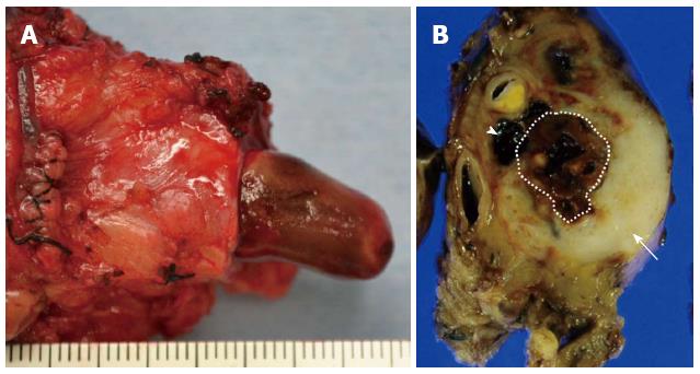
Figure 4 Pathological findings.
A: The dilated main pancreatic duct was filled with a papillary projecting tumor; B: The main tumor was dark reddish-brown (arrow head) and white (arrow) on the surface, and showed significant dilatation of the main pancreatic duct (dotted line region).
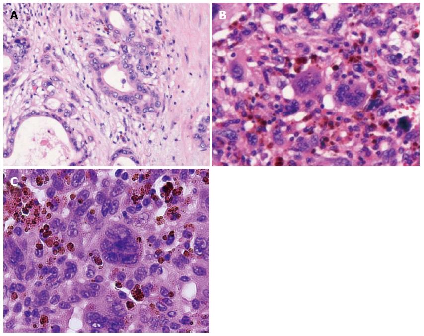
Figure 5 Microscopic appearance of the tumor.
Hematoxylin and eosin staining showed adenocarcinoma (tub1, tub2) in the white component, bizarre mono-and multi-nucleated giant cells in the dark reddish-brown component and a projecting tumor into the main pancreatic duct. A: the white component; B: the dark red-brown component; C: the projecting tumor.
- Citation: Okazaki M, Makino I, Kitagawa H, Nakanuma S, Hayashi H, Nakagawara H, Miyashita T, Tajima H, Takamura H, Ohta T. A case report of anaplastic carcinoma of the pancreas with remarkable intraductal tumor growth into the main pancreatic duct. World J Gastroenterol 2014; 20(3): 852-856
- URL: https://www.wjgnet.com/1007-9327/full/v20/i3/852.htm
- DOI: https://dx.doi.org/10.3748/wjg.v20.i3.852


