Copyright
©2014 Baishideng Publishing Group Co.
World J Gastroenterol. Jan 21, 2014; 20(3): 774-785
Published online Jan 21, 2014. doi: 10.3748/wjg.v20.i3.774
Published online Jan 21, 2014. doi: 10.3748/wjg.v20.i3.774
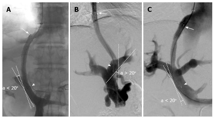
Figure 1 Stent position classifications.
The initial stent position was classified according to the angiography imaging as follows: A: Suboptimal in the hepatic vein (HV) (arrow) and optimal in the portal vein (PV) (arrow head); B: Optimal in the HV (arrow) and suboptimal in the PV (arrow head); C: Optimal in the HV (arrow) and optimal in the PV (arrow head).
Figure 2 Selection flowchart for the consecutive patients who underwent transjugular intrahepatic portosystemic shunt between March 2001 and July 2010.
TIPS: Transjugular intrahepatic portosystemic shunt.
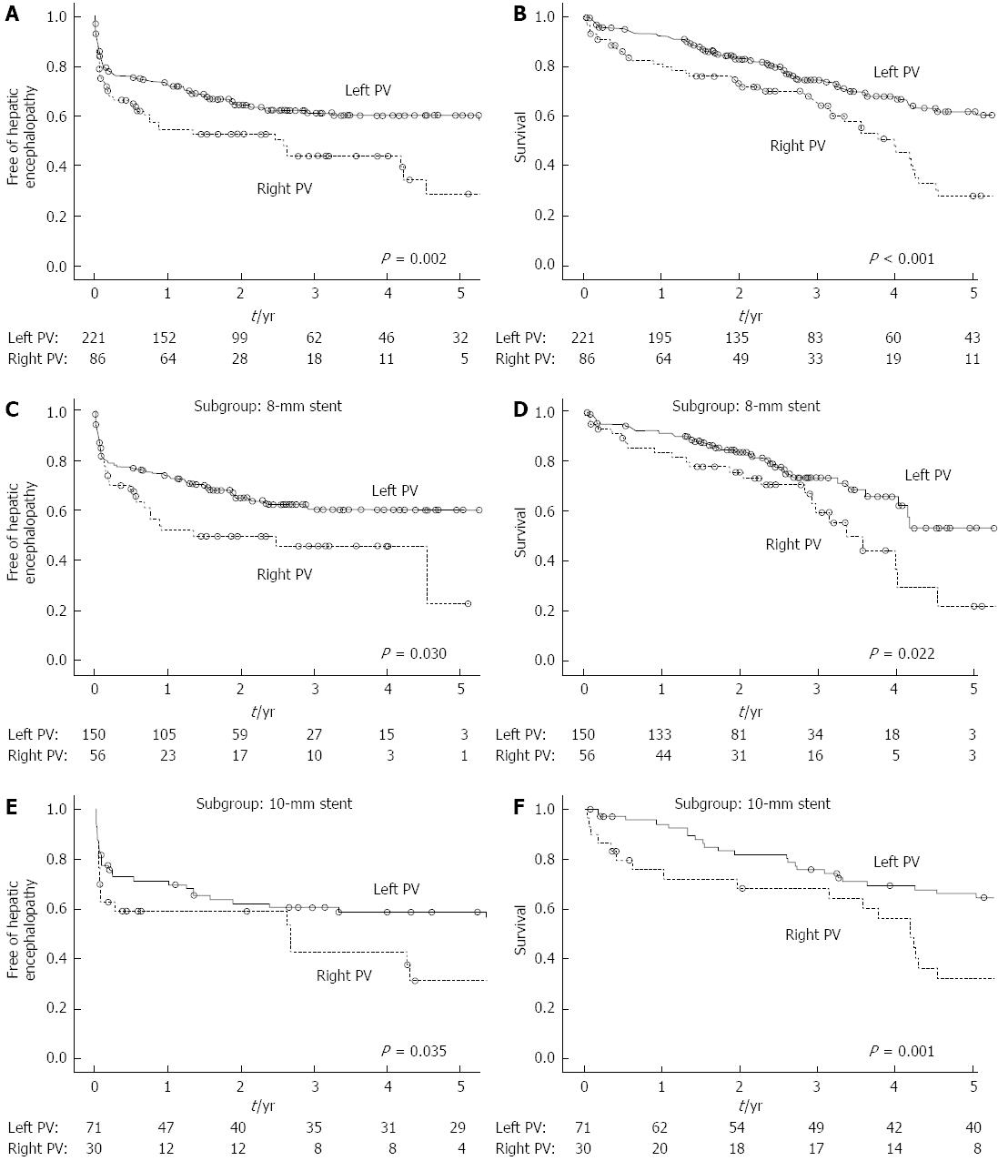
Figure 3 Hepatic encephalopathy results from the Kaplan-Meier analyses.
Comparison of hepatic encephalopathy between the patients with a transjugular intrahepatic portosystemic shunt (TIPS) to the left portal vein (PV) and those with a TIPS to the right PV in all patients (A), an 8-mm stent subgroup (C) and a 10-mm stent subgroup (E). Comparisons of survival between the patients with a TIPS to the left PV and those with a TIPS to the right PV in all patients (B), an 8-mm stent subgroup (D) and a 10-mm stent subgroup (F).
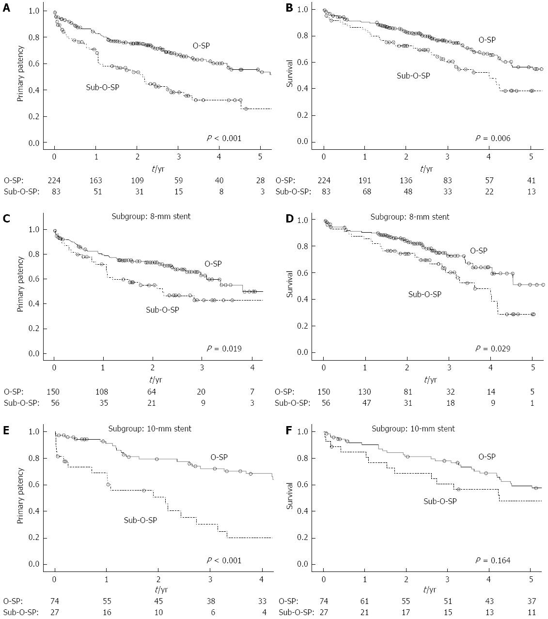
Figure 4 Patency results from the Kaplan-Meier analyses.
Comparison of primary patency between patients with optimal initial stent position (O-SP) and those with sub-O-SP in all patients (A), an 8-mm stent subgroup (C) and a 10-mm stent subgroup (E). Comparisons of survival between the patients with O-SP and those with sub-O-SP in all patients (B), an 8-mm stent subgroup (D) and a 10-mm stent subgroup (F).
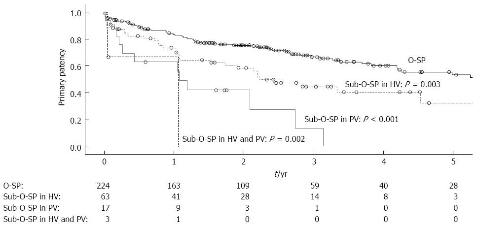
Figure 5 Patency results in patients with different stent positions.
Comparison of primary patency between patients with optimal initial stent position (O-SP) and those with sub-O-SP in the hepatic vein (HV) only, sub-O-SP in the portal vein (PV) only and sub-O-SP in both the HV and PV.
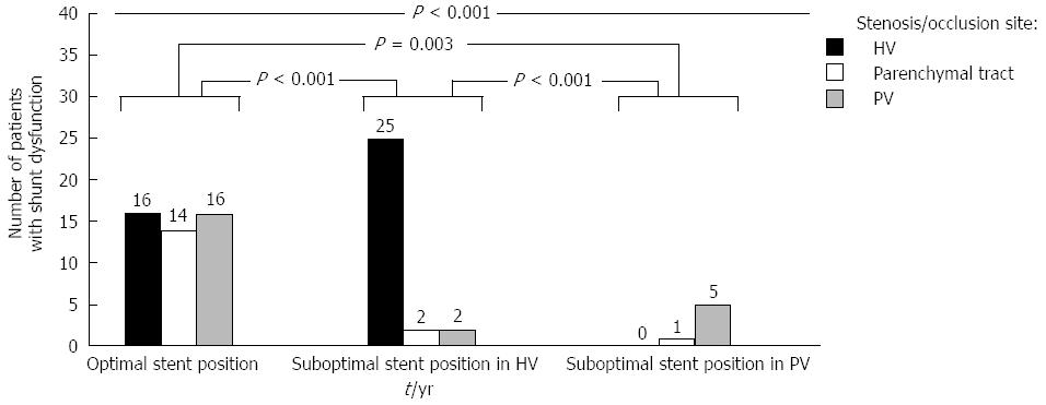
Figure 6 Relationship between stent positions and stenosis/occlusion sites.
The stenosis/occlusion sites of the first shunt dysfunction among patients with optimal initial stent position (O-SP) in the hepatic vein(HV) and portal vein (PV), patients with sub-O-SP in the HV and patients with sub-O-SP in the PV are significantly different.
- Citation: Bai M, He CY, Qi XS, Yin ZX, Wang JH, Guo WG, Niu J, Xia JL, Zhang ZL, Larson AC, Wu KC, Fan DM, Han GH. Shunting branch of portal vein and stent position predict survival after transjugular intrahepatic portosystemic shunt. World J Gastroenterol 2014; 20(3): 774-785
- URL: https://www.wjgnet.com/1007-9327/full/v20/i3/774.htm
- DOI: https://dx.doi.org/10.3748/wjg.v20.i3.774


