Copyright
©2014 Baishideng Publishing Group Co.
World J Gastroenterol. Jan 21, 2014; 20(3): 755-773
Published online Jan 21, 2014. doi: 10.3748/wjg.v20.i3.755
Published online Jan 21, 2014. doi: 10.3748/wjg.v20.i3.755
Figure 1 Results of immunohistochemical staining.
A-C: Immunohistochemistry for (A) Skp2, (B) Cks1, and (C) Ki67 proteins showed that stains were mainly expressed in the nuclei of cancer cells; D: The staining pattern of p27kip1 was somewhat heterogeneous. The pattern of nuclear staining was mainly noticed, and some of them were expressed as diffuse cytoplasmic staining. The staining intensity of Skp2, Cks1, and p27kip1 was classified as follows: 0, no staining; 1, weak; 2, moderate; 3, strong. The staining pattern of Ki-67 was nuclear and classified as positive or negative. At least 20 high-power fields will be randomly chosen, and 2000 cells will be always counted. Skp2: S-phase kinase-associated protein 2; Cks1: Cyclin-dependent kinases regulatory subunit 1.
Figure 2 Relationship among the immunostaining intensity for S-phase kinase-associated protein 2, Cyclin-dependent kinases regulatory subunit 1, p27kip1, and Ki-67 LI.
Ki-67 LI significantly increased as the staining intensity of Skp2 and Cks1 increased. Ki-67 LI showed no significant correlation with the staining intensity of p27kip1. Statistical analyses were performed by one-way ANOVA. bP < 0.01 between groups. Skp2: S-phase kinase-associated protein 2; Cks1: Cyclin-dependent kinases regulatory subunit 1.
Figure 3 Results of Kaplan-Meier survival analysis.
By Kaplan-Meier survival analysis, tumor size larger than 2 cm (A), advanced American Joint Commission on Cancer stage (B), tumor differentiation (C), R1 resection (D), angiolymphatic tumor invasion (E), and the intensity of Skp2 immunostaining (F) were significantly associated with overall survival in patients with extrahepatic cholangiocarcinoma. Skp2: S-phase kinase-associated protein 2.
Figure 4 MTT assay after the addition of exogenous epidermal growth factor.
Exogenous epidermal growth factor (EGF) (especially 0.1-10 ng/mL) significantly increased the proliferation indices of SNU-1196, SNU-1079, and SNU-245 cells. Statistical analyses were performed by one-way ANOVA. bP < 0.01 vs control. Data are representative of triplicate experiments.
Figure 5 Results of quantitative polymerase chain reaction for S-phase kinase-associated protein 2, Cyclin-dependent kinases regulatory subunit 1, and p27kip1 after the addition of exogenous epidermal growth factor in (A) SNU-1196, (B) SNU-1079, and (C) SNU-245 cells.
Data are representative of triplicate experiments. Statistical analyses were performed by one-way ANOVA. bP < 0.01 vs control. EGF: Exogenous epidermal growth factor; Skp2: S-phase kinase-associated protein 2; Cks1: Cyclin-dependent kinases regulatory subunit 1.
Figure 6 Results of western blotting for S-phase kinase-associated protein 2, Cyclin-dependent kinases regulatory subunit 1, and p27kip1 after the addition of exogenous epidermal growth factor in (A) SNU-1196, (B) SNU-1079, and (C) SNU-245 cells.
Data are representative of triplicate experiments. Skp2: S-phase kinase-associated protein 2; Cks1: Cyclin-dependent kinases regulatory subunit 1.
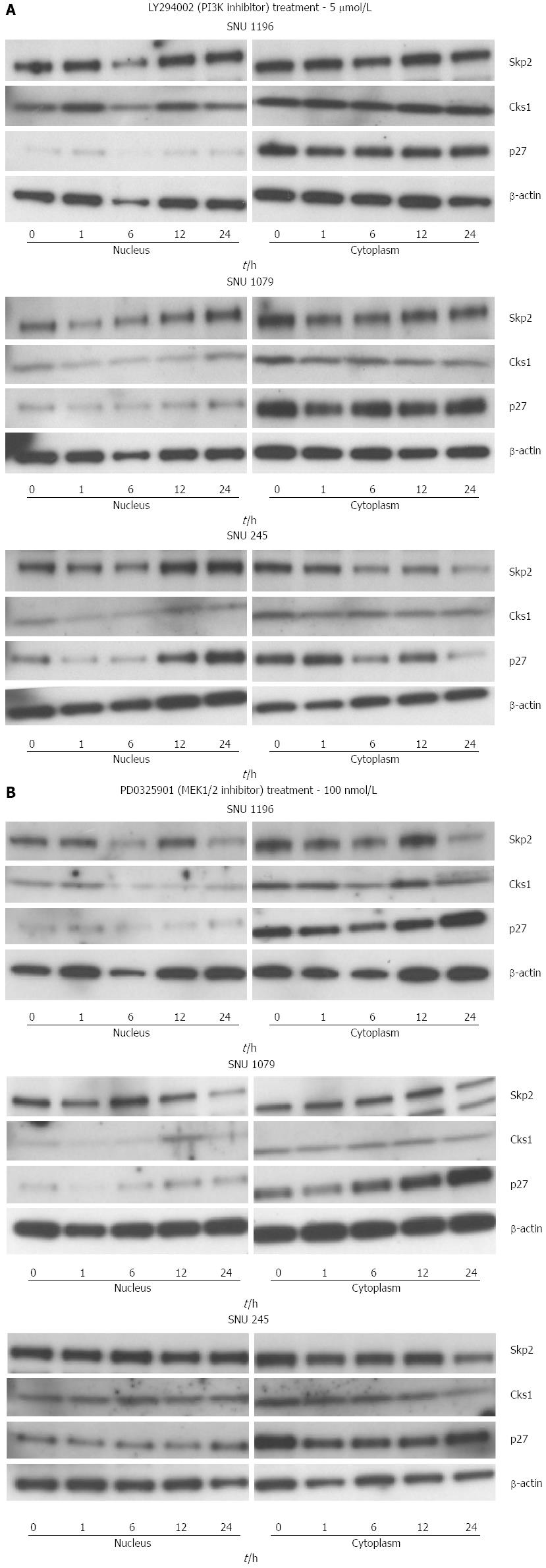
Figure 7 Effect of LY294002 (PI3K inhibitor, 5 μmol/L), and PD0325901 (MEK1/2 inhibitor, 100 nmol/L) on the protein levels of S-phase kinase-associated protein 2/Cyclin-dependent kinases regulatory subunit 1 and p27kip1 in SNU-1196, SNU-1079, and SNU-245 cells.
Data are representative of triplicate experiments. Skp2: S-phase kinase-associated protein 2; Cks1: Cyclin-dependent kinases regulatory subunit 1.
Figure 8 Binding of E2F1 transcription factor to the promoter region of Skp2 gene.
Chromatin immunoprecipitation assay shows the direct binding of E2F1 transcription factor to the promoter region of the Skp2 gene after the addition of 10 ng/mL of EGF in SNU-245, SUN-1196, SUN-1079, and NIH3T3 cells.
- Citation: Kim JY, Kim HJ, Park JH, Park DI, Cho YK, Sohn CI, Jeon WK, Kim BI, Kim DH, Chae SW, Sohn JH. Epidermal growth factor upregulates Skp2/Cks1 and p27kip1 in human extrahepatic cholangiocarcinoma cells. World J Gastroenterol 2014; 20(3): 755-773
- URL: https://www.wjgnet.com/1007-9327/full/v20/i3/755.htm
- DOI: https://dx.doi.org/10.3748/wjg.v20.i3.755


