Copyright
©2014 Baishideng Publishing Group Inc.
World J Gastroenterol. Aug 7, 2014; 20(29): 9850-9861
Published online Aug 7, 2014. doi: 10.3748/wjg.v20.i29.9850
Published online Aug 7, 2014. doi: 10.3748/wjg.v20.i29.9850
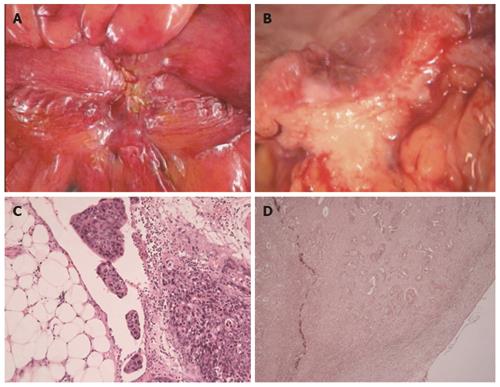
Figure 1 Macroscopic and microscopic features of peritoneal involvement.
A: Peritoneal puckering; B: Area with peritoneal puckering correlates with the invasive edge of the tumor on sectioning; C: Adenocarcinoma in a peritoneal cleft in a pT4a case; D: Invasion through peritoneal elastic lamina highlighted with an elastic stain.
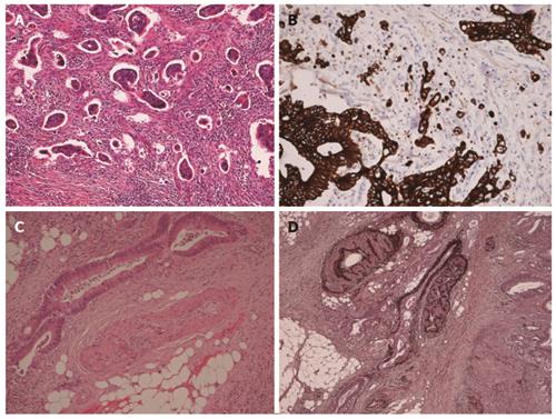
Figure 2 Histological appearances of tumor budding and vascular invasion.
A: Tumor budding; B: Cytokeratin immunohistochemical stain highlights tumor buds; C: “Orphan” artery sign - an elongated tumor profile is identified adjacent to an artery with no visible accompanying vein; D: Elastic stain highlights elastic fibres in the walls of arteries and an adjacent vein filled with tumor.
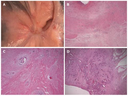
Figure 3 Pathological appearances of tumor regression.
A: Regressed tumor with the appearance of a mucosal scar; B: No residual tumor seen in tumor regression grading 1 (TRG1); C: Fibrosis outgrows tumor in TRG2; D: Extensive residual tumor in TRG3.
- Citation: Maguire A, Sheahan K. Controversies in the pathological assessment of colorectal cancer. World J Gastroenterol 2014; 20(29): 9850-9861
- URL: https://www.wjgnet.com/1007-9327/full/v20/i29/9850.htm
- DOI: https://dx.doi.org/10.3748/wjg.v20.i29.9850


