Copyright
©2014 Baishideng Publishing Group Inc.
World J Gastroenterol. Aug 7, 2014; 20(29): 10208-10211
Published online Aug 7, 2014. doi: 10.3748/wjg.v20.i29.10208
Published online Aug 7, 2014. doi: 10.3748/wjg.v20.i29.10208
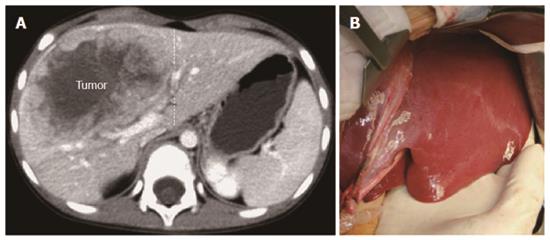
Figure 1 Cross-sectional imaging (A) and operative view (B) of the spatial relationship between the liver mass and the small left lateral section.
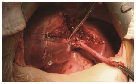
Figure 2 Right portal vein ligation and in-situ split between segment 4 and left lateral section.
A main branch of segment 4 portal pedicle was slung and later divided.
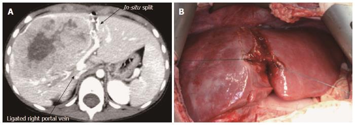
Figure 3 Volumetric changes induced by associating liver partition and portal vein ligation for stage hepatectomy.
A: Cross-sectional imaging on seventh postoperative day after in-situ split shows significant left lateral section hypertrophy; B: The operative view shows the dusky-looking segment 4 and the hypertrophied left lateral section.
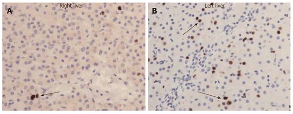
Figure 4 Immunohistochemical staining for Ki-67 (brown nuclei) showed the significant difference in cellular proliferative activity between the right liver (A) and left lateral section (B) after associating liver partition and portal vein ligation (arrows) for stage hepatectomy.
- Citation: Chan A, Chung PH, Poon RT. Little girl who conquered the "ALPPS''. World J Gastroenterol 2014; 20(29): 10208-10211
- URL: https://www.wjgnet.com/1007-9327/full/v20/i29/10208.htm
- DOI: https://dx.doi.org/10.3748/wjg.v20.i29.10208


