Copyright
©2014 Baishideng Publishing Group Inc.
World J Gastroenterol. Aug 7, 2014; 20(29): 10024-10037
Published online Aug 7, 2014. doi: 10.3748/wjg.v20.i29.10024
Published online Aug 7, 2014. doi: 10.3748/wjg.v20.i29.10024
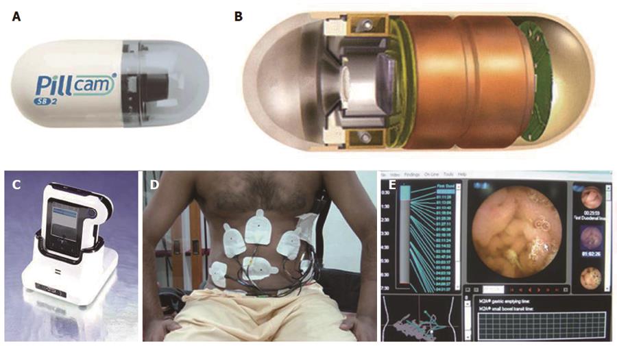
Figure 1 Components and design of capsule endoscopy system.
A: Capsule; B: Schematic diagram of components of capsule; C: Data recorder; D: Patient with sensor attachment; E: Dedicated work station.
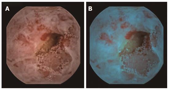
Figure 2 Ulcerated lesion at capsule endoscopy using chromoendoscopy.
A: White light; B: Blue light (with Fuji intelligent color enhancement).
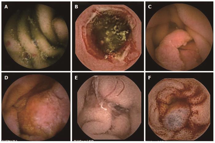
Figure 3 Lesions at capsule endoscopy seen in patients with obscure gastrointestinal bleed.
A: Arteriovenous malformation; B: Non-steroidal anti-inflammatory drugs ulcer; C: Polyp; D: Adenocarcinoma; E: Hook worm; F: Small intestinal varix.
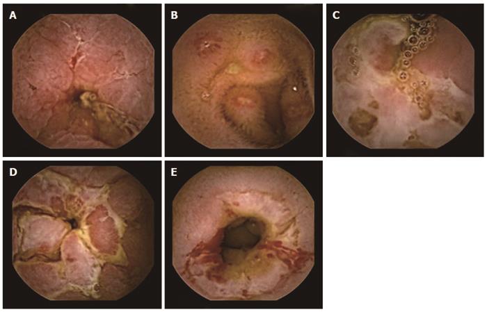
Figure 4 Capsule endoscopy in patients with Crohn’s disease.
A: Fissuring; B: Multiple aphthous ulcers; C: Serpiginous ulcer; D: Cobblestoning; E: Stricture.
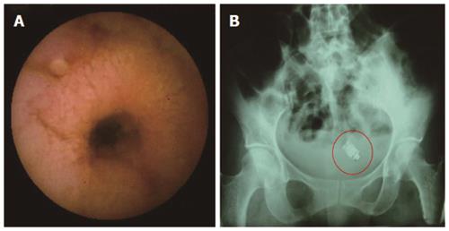
Figure 5 Capsule retention at stricture.
A: Stricture seen at capsule endoscopy; B: X-ray showing the retained capsule (red circle).
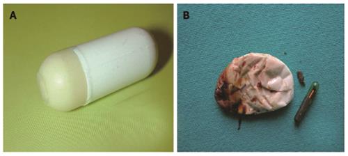
Figure 6 Agile patency capsule.
A: Before disintegration; B: After disintegration.
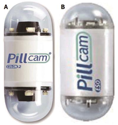
Figure 7 Pillcam.
A: Colonic capsule; B: Esophageal capsule.
- Citation: Goenka MK, Majumder S, Goenka U. Capsule endoscopy: Present status and future expectation. World J Gastroenterol 2014; 20(29): 10024-10037
- URL: https://www.wjgnet.com/1007-9327/full/v20/i29/10024.htm
- DOI: https://dx.doi.org/10.3748/wjg.v20.i29.10024


