Copyright
©2014 Baishideng Publishing Group Inc.
World J Gastroenterol. Jul 28, 2014; 20(28): 9570-9577
Published online Jul 28, 2014. doi: 10.3748/wjg.v20.i28.9570
Published online Jul 28, 2014. doi: 10.3748/wjg.v20.i28.9570
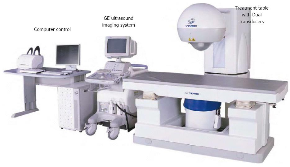
Figure 1 FEP-BY series high intensity focused ultrasound therapy system.
The system includes a computer control system, a GE ultrasound imaging system, and a treatment table with dual transducers.
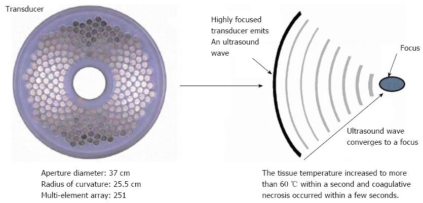
Figure 2 Principles of the high intensity focused ultrasound therapy system.
The tissue temperature increased to more than 60 °C within a second and coagulative necrosis occurred within a few seconds. The configuration (aperture diameter and radius of curvature) is for the structure used for reduction of the risk of skin burn.
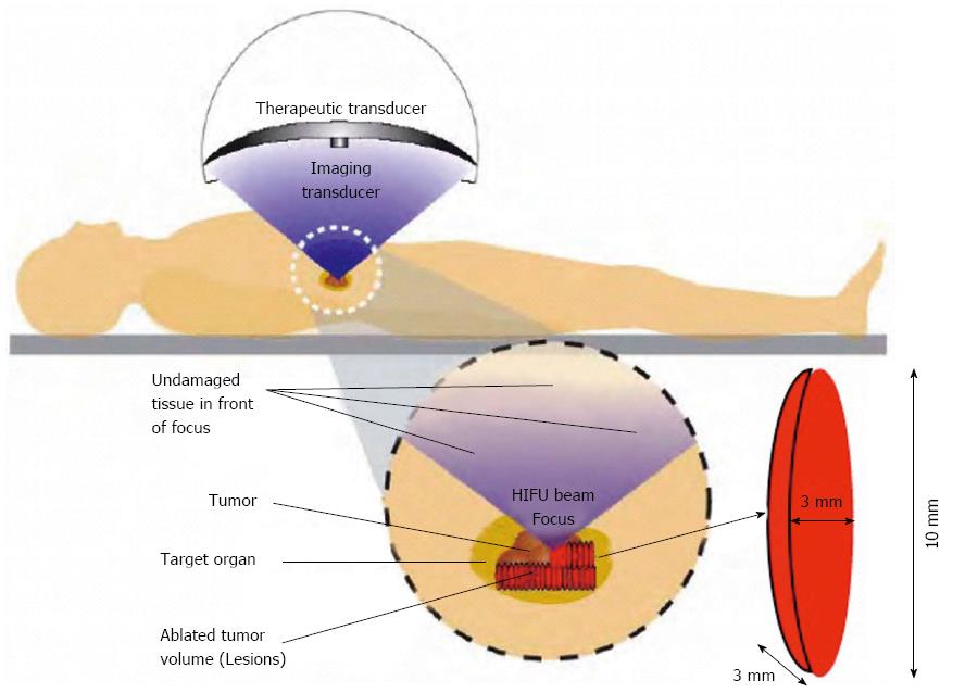
Figure 3 Principles of the high intensity focused ultrasound therapy system.
The FEP-BY Series High Intensity Focused Ultrasound (HIFU) therapy system has the ability to convey HIFU from an external source deep into tissues with a large convergence angle. The focus is oval in shape, with a short axis of approximately 3 mm and a long axis of 10 mm. Transducers have a multi-element array and concave focusing. The angle of convergence is 80°. The size of the focal point is smaller than 3 mm × 3 mm × 10 mm.
Figure 4 High intensity focused ultrasound therapy.
The system has 2 transducers, with the upper transducer being used for pancreatic cancer. Ultrasound gel was painted thoroughly over the target region. The position, size, and relationship of the tumor to the adjacent organ were determined by imaging ultrasonography before the therapy. During therapy, the patient lied supine on the treatment table.
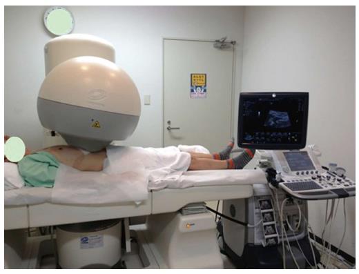
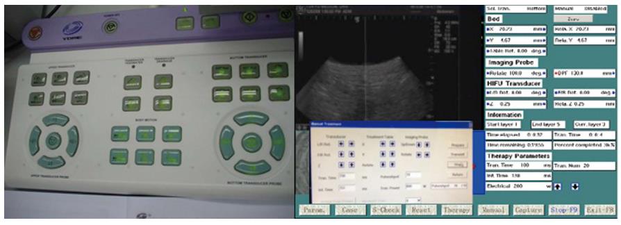
Figure 5 Monitoring system and treatment table.
The B-mode ultrasound was used to define the target area, therapeutic range, therapeutic layers, and power. The whole procedure of the treatment and the shift of focus were automatically controlled by computer.
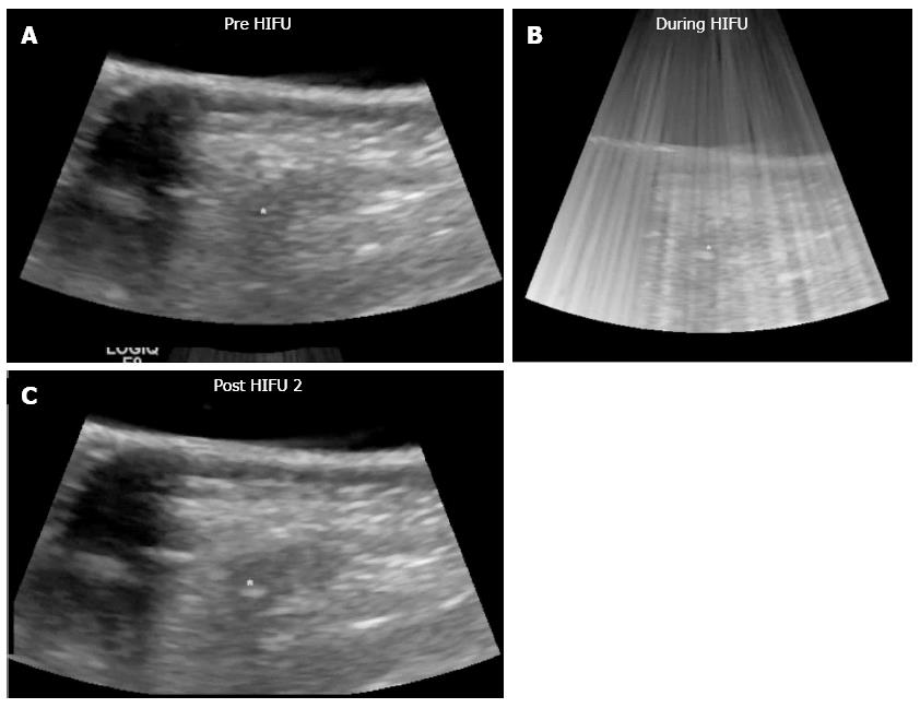
Figure 6 High intensity focused ultrasound therapy.
The whole procedure of the treatment and the focus steering were automatically controlled by computer. The yellow mark in the “Pre HIFU” image is the target location of the focal point. The echogenic region below the yellow mark is indicative of cavitation bubbles generated by the application of HIFU. HIFU: High Intensity Focused Ultrasound.
- Citation: Sofuni A, Moriyasu F, Sano T, Itokawa F, Tsuchiya T, Kurihara T, Ishii K, Tsuji S, Ikeuchi N, Tanaka R, Umeda J, Tonozuka R, Honjo M, Mukai S, Fujita M, Itoi T. Safety trial of high-intensity focused ultrasound therapy for pancreatic cancer. World J Gastroenterol 2014; 20(28): 9570-9577
- URL: https://www.wjgnet.com/1007-9327/full/v20/i28/9570.htm
- DOI: https://dx.doi.org/10.3748/wjg.v20.i28.9570


