Copyright
©2014 Baishideng Publishing Group Inc.
World J Gastroenterol. Jul 21, 2014; 20(27): 9055-9071
Published online Jul 21, 2014. doi: 10.3748/wjg.v20.i27.9055
Published online Jul 21, 2014. doi: 10.3748/wjg.v20.i27.9055
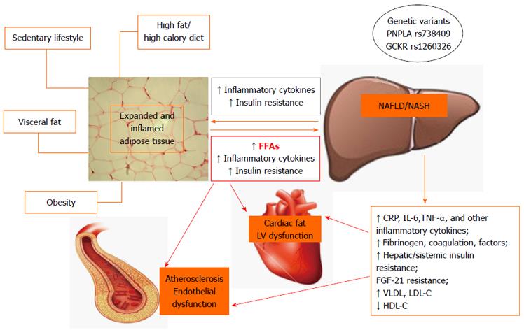
Figure 1 Suggested pathophysiological mechanisms linking nonalcoholic fatty liver disease to atherosclerosis and cardiac abnormalities in obese subjects.
FFAs: Fatty free acids; CRP: C-reactive protein; IL-6: Interleukin-6; TNF-α: Tumor necrosis factor-α; FGF-21: Fibroblast growth factor-21; VLDL: Very low density lipoprotein; LDL-c: Low density lipoprotein cholesterol; HDL-c: High density lipoprotein cholesterol; NAFLD: Nonalcoholic fatty liver disease; NASH: Nonalcoholic steatohepatitis.
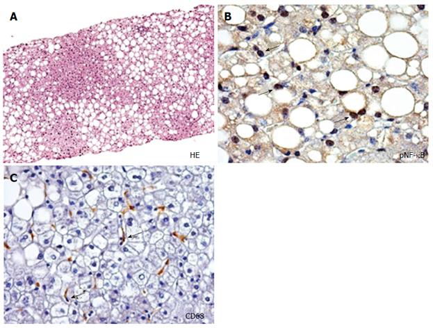
Figure 2 Histological and immunohistochemical features of nonalcoholic fatty liver disease.
A: Hematoxylin-Eosin (HE) in nonalcoholic fatty liver disease (NAFLD) biopsy shows hepatic steatosis (fatty liver). Original Magnification: 10 ×; B: Immunohistochemistry for phosphorylated (p) nuclear factor κB shows the nuclear expression by hepatocytes in NAFLD (arrows). Original Magnification: 40 ×; C: Macrophages in NAFLD. Immunohistochemistry for CD68 shows the presence of macrophages in nonalcoholic steatohepatitis (yellow arrows). Original Magnification: 40 ×. Photos were obtained from a liver biopsy of a 60-year-old male affected by NAFLD. Photos are original and taken in Prof. Gaudio’s Laboratory.
- Citation: Pacifico L, Chiesa C, Anania C, Merulis AD, Osborn JF, Romaggioli S, Gaudio E. Nonalcoholic fatty liver disease and the heart in children and adolescents. World J Gastroenterol 2014; 20(27): 9055-9071
- URL: https://www.wjgnet.com/1007-9327/full/v20/i27/9055.htm
- DOI: https://dx.doi.org/10.3748/wjg.v20.i27.9055


