Copyright
©2014 Baishideng Publishing Group Inc.
World J Gastroenterol. Jul 14, 2014; 20(26): 8726-8728
Published online Jul 14, 2014. doi: 10.3748/wjg.v20.i26.8726
Published online Jul 14, 2014. doi: 10.3748/wjg.v20.i26.8726
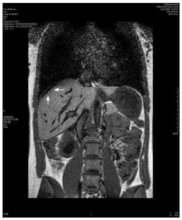
Figure 1 T2 weighted coronal magnetic resonance imaging image of subcapsular liver hematoma.
The arrows indicate a hyperdense area suggestive of the subcapsular liver hematoma.
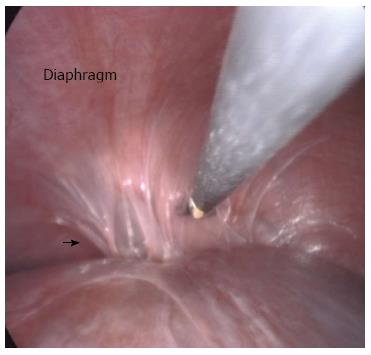
Figure 2 Laparoscopy image of perihepatic adhesions.
The arrow indicates the location of the perihepatic adhesions to the diaphragm that were found during laparoscopy.
- Citation: Koeneman MM, Koek GH, Bemelmans M, Peeters LL. Perihepatic adhesions: An unusual complication of hemolysis, elevated liver enzymes and low platelet syndrome. World J Gastroenterol 2014; 20(26): 8726-8728
- URL: https://www.wjgnet.com/1007-9327/full/v20/i26/8726.htm
- DOI: https://dx.doi.org/10.3748/wjg.v20.i26.8726


