Copyright
©2014 Baishideng Publishing Group Inc.
World J Gastroenterol. Jul 7, 2014; 20(25): 8320-8324
Published online Jul 7, 2014. doi: 10.3748/wjg.v20.i25.8320
Published online Jul 7, 2014. doi: 10.3748/wjg.v20.i25.8320
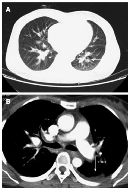
Figure 1 Chest computed tomography scaning.
A: Low density lesion (arrow) occurs along the pulmonary vessels; B: Small effusion (arrow) in the left pleural cavity.
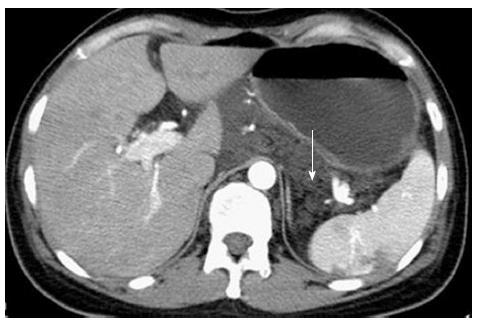
Figure 2 Abdominal computed tomography reveals multiple small cystic lesions (arrow) without enhancement in the abdominal cavity.
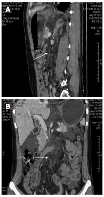
Figure 3 Computed tomography enterograph reveals multiple small cystic lesions (arrow) without enhancement in the abdominal cavity.
A: Sagittal images; B: Coronal images.
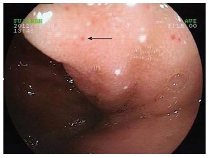
Figure 4 Single balloon enteroscopy reveals duodenal lymphangiectasia with hemorrhage (arrow).
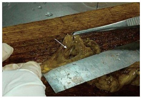
Figure 5 Broken vesica of the gross specimen (arrow).
- Citation: Lin RY, Zou H, Chen TZ, Wu W, Wang JH, Chen XL, Han QX. Abdominal lymphangiomatosis in a 38-year-old female: Case report and literature review. World J Gastroenterol 2014; 20(25): 8320-8324
- URL: https://www.wjgnet.com/1007-9327/full/v20/i25/8320.htm
- DOI: https://dx.doi.org/10.3748/wjg.v20.i25.8320


