Copyright
©2014 Baishideng Publishing Group Inc.
World J Gastroenterol. Jul 7, 2014; 20(25): 8292-8297
Published online Jul 7, 2014. doi: 10.3748/wjg.v20.i25.8292
Published online Jul 7, 2014. doi: 10.3748/wjg.v20.i25.8292
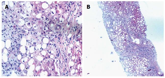
Figure 1 Macrovesicular steatosis (A) and steatosis and cirrhotic nodules (B).
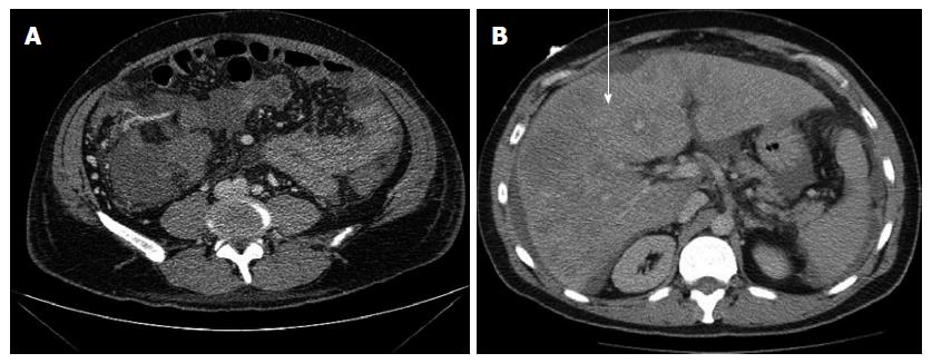
Figure 2 Contrast computed tomography abdomen (arrow).
The liver consistent with cirrhosis (arrow).
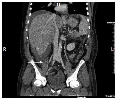
Figure 3 CTA and CTV of abdomen (arrow).
This revealed right abdominal mesenteric varices with a focal area of more consolidated abnormal enhancement in the right lower quadrant (arrow).
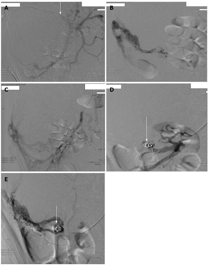
Figure 4 Superior mesenteric (A) portal venogram (B), 8- coil embolization of colonic branch (C, D) and cecal branch (E) of superior mesenteric vein (arrow).
- Citation: Edula RG, Qureshi K, Khallafi H. Hemorrhagic ascites from spontaneous ectopic mesenteric varices rupture in NASH induced cirrhosis and successful outcome: A case report. World J Gastroenterol 2014; 20(25): 8292-8297
- URL: https://www.wjgnet.com/1007-9327/full/v20/i25/8292.htm
- DOI: https://dx.doi.org/10.3748/wjg.v20.i25.8292


