Copyright
©2014 Baishideng Publishing Group Inc.
World J Gastroenterol. Jul 7, 2014; 20(25): 8253-8259
Published online Jul 7, 2014. doi: 10.3748/wjg.v20.i25.8253
Published online Jul 7, 2014. doi: 10.3748/wjg.v20.i25.8253
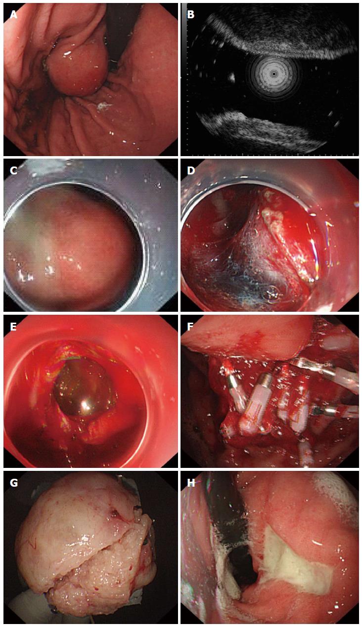
Figure 1 Endoscopic full-thickness resection treatment of gastric stromal tumors arising from the muscularis propria.
A: A protruding submucosal lesion in the gastric body; B: Endoscopic ultrasound showing that the lesion arose from the muscularis propria; C: Submucosal injection of saline containing adrenaline and indigo carmine; D: Application of the IT knife to isolate the stromal tumor along its periphery; E: An “artificial perforation” observed after stromal tumor resection, sealed using titanium clips; F: Sealing of the perforation with multiple titanium clips; G: Resected tumor with the mucosa removed (5 cm in diameter); H: View 72 d after the operation, showing that the perforation healed well, with only ulcer residue remaining.
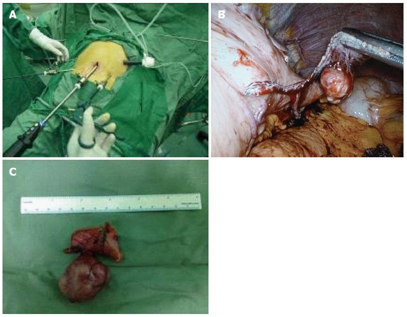
Figure 2 Laparoscopic resection of gastric stromal tumors.
A: Layout of instruments for laparoscopic surgery; B: Laparoscopic resection of gastric stromal tumor; C: Removed tumor (4 cm in diameter).
- Citation: Huang LY, Cui J, Wu CR, Zhang B, Jiang LX, Xian XS, Lin SJ, Xu N, Cao XL, Wang ZH. Endoscopic full-thickness resection and laparoscopic surgery for treatment of gastric stromal tumors. World J Gastroenterol 2014; 20(25): 8253-8259
- URL: https://www.wjgnet.com/1007-9327/full/v20/i25/8253.htm
- DOI: https://dx.doi.org/10.3748/wjg.v20.i25.8253


