Copyright
©2014 Baishideng Publishing Group Inc.
World J Gastroenterol. Jun 21, 2014; 20(23): 7424-7433
Published online Jun 21, 2014. doi: 10.3748/wjg.v20.i23.7424
Published online Jun 21, 2014. doi: 10.3748/wjg.v20.i23.7424
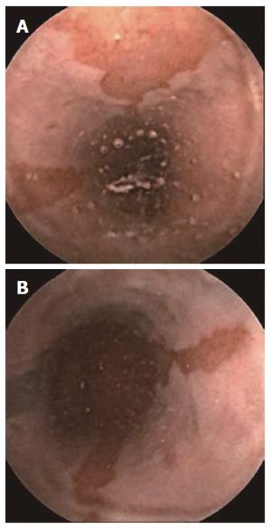
Figure 1 Suspected Barrett’s esophagus seen with PillCam ESO.
A: Suspected long Barrett’s esophagus; B: Ectopic tissue mucosa ascending from Z line.
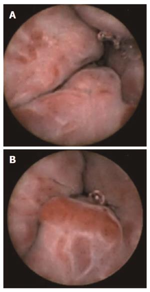
Figure 2 Esophageal varices seen with PillCam ESO.
A: Large esophageal varices with red spots; B: Large esophageal varices in distal esophagus.
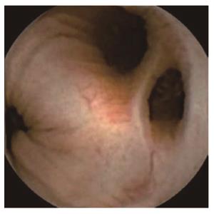
Figure 3 Diverticulosis coli.
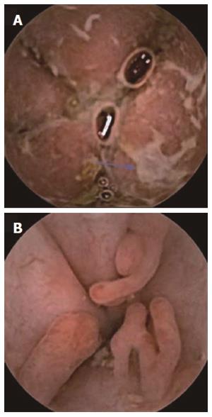
Figure 4 Severe (A) and pseudopolyps in inactive (B) ulcerative colitis.
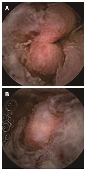
Figure 5 Colorectal cancer.
A: Ulcerated sigmoid neoformation; B: Partially stenosing sigmoid neoformation.
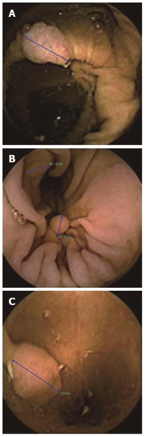
Figure 6 Polyps.
PillCam Colon-2 using polyp size estimation tool. PillCam Colon-2 using polyp size estimation tool. A: Senile ascending colon polyp; B: Millimetric descending colon polyps; C: Semi-pediculated polyp greater than 1cm in the sigmoid.
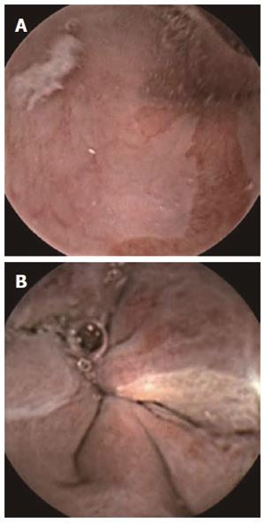
Figure 7 Suspected Barrett’s esophagus (A) and esophageal varices seen (B) with PillCam Colon.
- Citation: Romero-Vázquez J, Argüelles-Arias F, García-Montes JM, Caunedo-Álvarez &, Pellicer-Bautista FJ, Herrerías-Gutiérrez JM. Capsule endoscopy in patients refusing conventional endoscopy. World J Gastroenterol 2014; 20(23): 7424-7433
- URL: https://www.wjgnet.com/1007-9327/full/v20/i23/7424.htm
- DOI: https://dx.doi.org/10.3748/wjg.v20.i23.7424


